Why Might I Need A Breast Ultrasound
A breast ultrasound is most often done to find out if a problem found by a mammogram or physical exam of the breast may be a cyst filled with fluid or a solid tumor.
Breast ultrasound is not usually done to screen for breast cancer. This is because it may miss some early signs of cancer. An example of early signs that may not show up on ultrasound are tiny calcium deposits called microcalcifications.
Ultrasound may be used if you:
-
Have particularly dense breast tissue. A mammogram may not be able to see through the tissue.
-
Are pregnant. Mammography uses radiation, but ultrasound does not. This makes it safer for the fetus.
-
Are younger than age 25
Your healthcare provider may also use ultrasound to look at nearby lymph nodes, help guide a needle during a biopsy, or to remove fluid from a cyst.
Your healthcare provider may have other reasons to recommend a breast ultrasound.
What Do Cysts Look Like On An Ultrasound
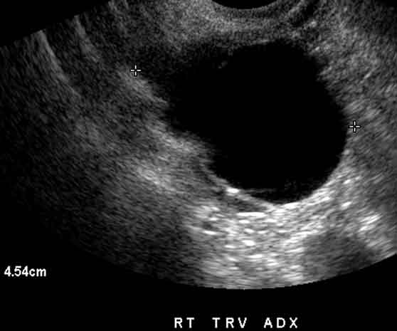
A simple cyst typically is round or oval, anechoic, and has smooth, thin walls. It contains no solid component or septation , and no internal flow is visible on color Doppler imaging. Cyst < 3. cm: No action necessary the cyst is a normal physiologic finding and should be referred to as a follicle …
Male Breast Ultrasound: Gynecomastia Versus Breast Cancer
Male breast cancer is very rare, but one condition, gynecomastia, which is the development of abnormally large breasts in men, is quite common. Gynecomastia is usually caused by excessive growth of fibroglandular breast tissue in men in their 60s or as the result of hormonal imbalances.
However, in rare cases, breast cancer can be the cause of gynecomastia so, a full mammographic investigation is always necessary.
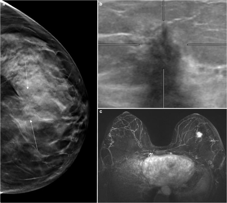
In the mammogram below, one can see the increase in the density of the fibroglandular tissues behind the nipple. It appears to be developing in a concentric pattern. The contour of the dense area is concave-outward and interspersed with fat.
There are no well-formed masses and no suspicious microcalcifications. It certainly would appear to be gynecomastia from natural or hormonal causes and not breast cancer.
In the sonogram of the same breast, one notes that the density appears hypoechoic with ill-defined margins. The amount of tissue appears to be thicker than average in a male but the only way to know if anything abnormal is going on in the sonogram would be to compare it with an ultrasound of the other breast to see if the densities are about the same.
Read Also: How To Test For Male Breast Cancer
Breast Cancer Ultrasound Images
Next to heart disease, breast cancer is the leading cause of death in women all over the world. Although the exact cause of breast cancer is unclear, early detection and treatment saves lives and reduces cost. Fortunately, it is becoming easier to diagnose the disease through different technologies, with ultrasound imaging as one of the frequently used diagnostic tools.Here we show somebreast cancer ultrasound images and important facts for your information.
Lets Do Some Q& a About Mammography

How are mammograms done?
During a mammogram, your breasts are compressed between two firm surfaces to spread out the breast tissue. Then, an x-ray captures black and white images of your breasts that are displayed on a computer screen and examined by a doctor who looks for signs of cancer.
How can mammograms be used?
A mammogram can be used either for screening or for diagnostic purposes.
How often should you have a mammogram?
It all depends on your age and your risk of breast cancer.
How do I know when I should begin screening mammography?
Some general guidelines for when to begin screening mammography include women with an average risk of breast cancer and woman with a high risk of breast cancer.
What are the risks?
Some known risks and limitations of mammograms include the following: Mammograms:-
- expose you to low-dose radiation
- are not always accurate
- can be difficult to interpret in younger women
- may lead to additional testing
- can not detect all breast cancers
- may show cancer, but not all of the tumors can be cured
How do I prepare for my mammogram appointment?
- Choose a certified mammogram facility
- schedule the test for a time when your breasts are least likely to be tender
- bring your prior mammogram images
- do not use deodorant before your mammogram
- consider an over-the-counter pain medication if you find that having a mammogram is uncomfortable.
What can a radiologist possibly find on my mammogram imaging?
Well, possible findings can include:-
- calcium deposits
Also Check: Can Breast Cancer Be Inherited From Father’s Side
Breast Cancer Tumor Cells
Under the microscope, breast cancer cells may appear similar to normal breast cells. They also may look quite different, depending on the tumor’s growth and grade.
Cancer cells differ from normal cells in many ways. The cells may be arranged in clusters. They also may be seen invading blood vessels or lymphatic vessels.
The nucleus of cancer cells can be striking, with nuclei that are larger and irregular in shape. These centers will stain darker with special dyes. Often, there are extra nuclei rather than just one center.
Various Biopsy Methods For The Breast
Image-guided breast biopsy is currently the gold standard for thepathologic evaluation of breast cancer. It can be performed safely and reliablywith minimal invasiveness in clinical practice and with increased patientconvenience and decreased cost . Ultrasound, stereotactic mammography, magnetic resonanceimaging , and positron emission mammography are now successfully usedfor the guidance of the biopsy needle in order to obtain a proper tissue samplethat can be histologically assessed. The choice of image guidance for biopsy isbased on a variety of factors, including which modality best visualizes thelesion, the physicians clinical experience, patient comfort, cost, easeof access, and equipment availability. The common methods of biopsy includefine-needle aspirate biopsy, vacuum-assisted biopsy, and core-needle biopsy.Meta-analysis for various diagnostic biopsy methods for women at average risk ofcancer showed that SE estimates were higher than 0.90 and SP estimates werehigher than 0.91 for all methods .
Also Check: Can You Have Breast Cancer In Both Breast
Distinguishing Benign Masses From Malignant Masses
Originally, ultrasonography was primarily used to distinguish simple cysts, which did not require sampling, from solid masses that were usually examined with biopsy. In many cases, the results of these biopsies were benign. Improving equipment and scanning techniques have helped expand the applications of breast US. Linear-array high-frequency transducers are generally used.
Automated Whole Breast Ultrasound
Whole breast ultrasound exams are most often performed with a hand-held ultrasound device . The quality of the image can vary greatly, depending on the skill and experience of the person doing the exam.
Automated whole breast ultrasound is under study to see if it may help improve image quality . However, its not widely available or routinely used.
Read Also: What Is Survival Rate For Stage 4 Breast Cancer
Breast Ultrasound: Criteria For Benign Lesions
Several studies have described the sonographic characteristics commonly seen in benign lesions of the breast:
Scar Tissue Can Often Appear Suspicious
The image below contains a lesion with irregular, spiculated There does not appear to be a central mass to this lesion, which right away makes it less likely to be breast cancer. However, something this suspicious would likely need a biopsy to find out exactly what is going on. This lesion is more likely to be either a post-surgical scar or possibly a radial scar. In actual fact this particular image was taken from a woman who had breast surgery, so a post-surgical scar is the most probable diagnosis.
A closer look via magnification of the same lesion reveals a central radio-transparency likely caused by fat necrosis, and there is no central mass.
The spiculations around the lesion are likely a desmoplastic reaction to the surgery. .
Recommended Reading: What Does Stage 3 Triple Negative Breast Cancer Mean
Examples Of Breast Cancer Screening Mammography Interpretation
Even with ill-defined borders and spiculated margins, other factors make breast cancer an unlikely diagnosis
The X-ray image below shows a suspect breast mass of about 1 cm in diameter. Some architectural distortion is also apparent. An ultrasound image of the same lesion suggests that the lesion is solid. The mass appears to be hypoechoic with ill-defined, spiculated, and microlobulated margins.
It is not possible to rule out malignancy here because posterior acoustic shadowing is not present. When a lesion is homogeneous, good through-transmission of the ultrasound beam is possible, and malignant breast cancer lesions are typically not so homogeneous.
Characteristics Of Malignant Lesions

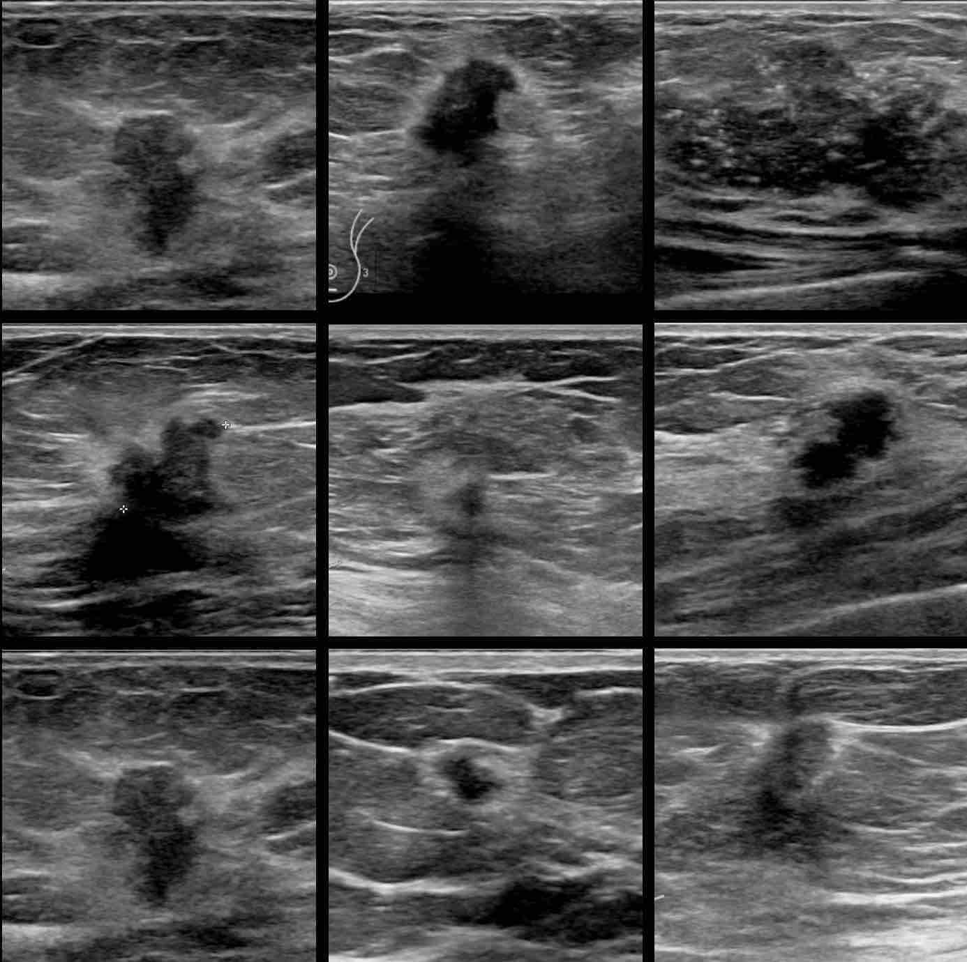
Malignant lesions are commonly hypoechoic lesions with ill-defined borders. Typically, a malignant lesion presents as a hypoechoic nodular lesion, which is taller than broader and has spiculated margins, posterior acoustic shadowing and microcalcifications . Three-dimensional scanners with the capability of reproducing high-resolution images in the coronal plane provide additional important information. The spiky extensions along the tissue planes can be well seen in coronal images .B]. It was initially believed that color Doppler scanning would add to the specificity of USG examination, but this has not proven to be very efficacious however, in certain situations it does help resolve the issue, particularly when there is significant vascularity present within highly cellular types of malignancies .
Malignant lesions. Transverse scan shows a typical malignant nodule that is taller than wide, with hypoechoic echotexture. Arrowheads indicate irregular spiculated margins. Some of the nodules may reveal a branching pattern . Sagittal view shows a nodule with multilobulated margins the presence of more than 34 lobulations is suspicious for malignancy. Sagittal and transverse scans show duct extension . M indicates the primary site. Duct extension appears smooth in outline in cross-section . Transverse scan shows a typical malignant lesion with irregular spiky margins, microcalcifications and a branching pattern. This lesion is classifiable as US-BIRADS category 4
Don’t Miss: Does Lung Cancer Metastasis To Breast
What Does The Equipment Look Like
Ultrasound machines consist of a computer console, video monitor and an attached transducer. The transducer is a small hand-held device that resembles a microphone. Some exams may use different transducers during a single exam. The transducer sends out inaudible, high-frequency sound waves into the body and listens for the returning echoes. The same principles apply to sonar used by boats and submarines.
The technologist applies a small amount of gel to the area under examination and places the transducer there. The gel allows sound waves to travel back and forth between the transducer and the area under examination. The ultrasound image is immediately visible on a video monitor. The computer creates the image based on the loudness , pitch , and time it takes for the ultrasound signal to return to the transducer. It also considers what type of body structure and/or tissue the sound is traveling through.
How Does A Radiologist See Breast Cancer On Mammography & Ultrasound
When you look at mammography or ultrasound images, you might wonder how radiologists make any sense of them. How can they identify potential cancers in those Rorschach tests of gray and white? While even the most advanced imaging technology doesnt allow radiologists to identify cancer with certainty, it does give them some strong clues about what deserves a closer look. Today well discuss a few things that radiologists are on the lookout for when examining mammography and breast ultrasound images.
When radiologists look at a mammogram, theyre looking for three primary things:
- Changes from what is seen in previous images
If youve had a mammogram before, it is helpful to give your current radiologist access to your previous mammography images. Anytime you visit a new mammography clinic, let them know where youve had breast imaging done in the past so they are able to note any changes over time.
Masses comprise a variety conditions, including cysts, benign solid tumors, and malignancies. Their size, shape, borders, and internal composition can give insight into whether they represent cancer. Cancerous tumors often appear as white masses with blurry or spiked borders, which indicate infiltration into the surrounding tissue. Cysts are often indistinguishable from solid tumors on a mammogram, so ultrasound is often used to determine whether a mass is solid or fluid filled .
Recommended Reading: Why Is Triple Negative Breast Cancer So Aggressive
Can A Radiologist Diagnose Breast Cancer From An Ultrasound
Dr. Kella: Breast radiologists specialize in interpreting images of the breast in order to diagnose and help treat different medical conditions of the breast. They read mammograms, breast ultrasounds, and breast MRIs, and perform diagnostic breast procedures that can help to diagnose and treat breast cancer.
Lactating Adenomas In Mammogram Images
Breast cancer is very uncommon in younger women. So, if a young woman who is pregnant came in for a screening of a palpable breast lump it is far more likely that the lesion is a fibroadenoma of some kind.
One common variation of fibroadenoma in pregnant women is a lactating adenoma, which is essentially a tubular adenoma that occurs in pregnant women. Lactating adenoma features the accumulation of milk secretions in addition to hyperplasia.
Breast X-rays are not normally given to pregnant women. Given that breast cancer is very unlikely and lactating adenoma is quite likely, ultrasound and possibly a fine needle aspiration biopsy would typically be utilized for diagnostic investigations.
The main concern with a lactating adenoma from the perspective of breast cancer is that the condition can occur simultaneously with breast cancer. However, on their own, they indicate no increase in the risk for subsequent breast cancer development.
In the ultrasound image of lactating adenoma below, one notes a hypoechoic, non-cystic mass in an ovoid shape. It has a long axis running parallel to the skin, posterior acoustic enhancement, and well-defined margins.
You May Like: What Causes Estrogen Positive Breast Cancer
When Should I Call My Doctor
- Feel a new or changing lump, dimpling, or other changes in your breast or armpit that are unusual for you.
- Have any nipple discharge, new inversion or skin changes of the nipple.
- Think a breast implant has ruptured.
A note from Cleveland Clinic
A breast ultrasound is a safe, painless test to examine targeted areas of breast tissue. Breast ultrasound provides detailed images of breast tissue and can help your provider diagnose breast cysts or lumps. For women with dense breasts, mammography is still the best screening tool. If you have dense breasts or a family history of breast cancer, ask your provider about scheduling a risk assessment with a clinical breast specialists and supplemental screening tools such as MRI and tomosythesis mammography.
Last reviewed by a Cleveland Clinic medical professional on 03/30/2021.
References
Treatment Planning Surgery And Posttreatment Follow
Berg et al showed the possible benefit of combining preoperative whole-breast US with mammography when breast-conservation surgery is planned. US demonstrated additional sites of multifocal and multicentric carcinoma, facilitating preoperative planning.
Several investigators have studied the role of US in the assessment of axillary lymph nodes for tumor involvement. Normal lymph nodes usually have a prominent echogenic fatty hilum and a thin hypoechoic cortex. Lymph nodes that lack a fatty echogenic hilum or are heterogeneous are considered suspicious. The appearances on US of benign and malignant lymph nodes overlap therefore, US-guided fine-needle aspiration biopsy of suspicious lymph nodes has been advocated. Krishnamurthy et al found that in approximately 12% of cases, false-negative results occur with US-guided axillary lymph node FNAB.
Deurloo et al showed that US-guided axillary lymph node FNAB reduces the number of the more time-consuming sentinel-node biopsy procedures that are needed.
Intraoperative US may be used to localize breast masses. It obviates the need for preoperative needle localization, offers more flexibility in choosing the incision site than preoperative needle localization, and may allow assessment of the tumor’s extent. However, intraoperative US is operator dependent, and as with breast needle localization, it may not help in localizing the carcinoma.
You May Like: How To Survive Stage 4 Breast Cancer
How Painful Is A Breast Biopsy
You will be awake during your biopsy and should have little discomfort. Many women report little pain and no scarring on the breast. However, certain patients, including those with dense breast tissue or abnormalities near the chest wall or behind the nipple, may be more sensitive during the procedure.
Is Breast Cancer Painless

This is an unfortunate case with a difficult initial diagnosis. Breast cancers are usually painless. She was due for further assessment TRO malignancy. Due to economic reason, she defaulted follow up and by the time she came back, the mass has grown from 5mm to 3cm. her GP is now arranging for surgical referral and biopsy.
Also Check: Where To Go To Get Checked For Breast Cancer
The Development Of Novel Ultrasound Technology Will Help Radiologists Differentiate Cancer From Benign Masses Preventing Invasive And Unnecessary Breast Biopsies And Multi
Breasts come in all shapes and sizes, as well as various densitiesan important consideration when screening for cancer. Nearly half of all patients have dense breasts, which make it more challenging to spot cancer in mammogram images. Ultrasound screening and follow-up can be better at catching breast cancer early in those with dense breast tissue. However, this imaging technique has a high false-positive rate, sometimes resulting in patients undergoing invasive aspiration procedures, unnecessary biopsies, and follow-up monitoring that requires some patients to wait up to two years for a definitive diagnosis.
New research to improve ultrasound techniques for breast cancer screening and diagnosis is on the way. Muyinatu Bell, the John C. Malone Associate Professor of Electrical and Computer Engineering, is leading a project to develop ultrasound technology that can help radiologists detect early-stage breast cancers, regardless of a patient’s breast tissue density. Bell leads the Whiting School of Engineering’s .
Image caption: Muyinatu Bell
Image credit: Will Kirk / Johns Hopkins University
When breast cancer is detected early, the chances of survival are very high almost 99% of patients diagnosed at the earliest stage live for five years or more, according to the CDC.
Not all breast lumps are cancerous. In fact, many lumps are fluid-filled masses, or cysts, which are usually benign. Other lumps are solid masses, or tumors that may be cancerous and warrant more analysis.