Scar Tissue After Breast Biopsy
Ask U.S. doctors your own question and get educational, text answers â its anonymous and free!
Ask U.S. doctors your own question and get educational, text answers â its anonymous and free!
HealthTap doctors are based in the U.S., board certified, and available by text or video.
Recommended Reading: Level 3 Breast Cancer
Lets Do Some Q& a About Mammography
How are mammograms done?
During a mammogram, your breasts are compressed between two firm surfaces to spread out the breast tissue. Then, an x-ray captures black and white images of your breasts that are displayed on a computer screen and examined by a doctor who looks for signs of cancer.
How can mammograms be used?
A mammogram can be used either for screening or for diagnostic purposes.
How often should you have a mammogram?
It all depends on your age and your risk of breast cancer.
How do I know when I should begin screening mammography?
Some general guidelines for when to begin screening mammography include women with an average risk of breast cancer and woman with a high risk of breast cancer.
What are the risks?
Some known risks and limitations of mammograms include the following: Mammograms:-
- expose you to low-dose radiation
- are not always accurate
- can be difficult to interpret in younger women
- may lead to additional testing
- can not detect all breast cancers
- may show cancer, but not all of the tumors can be cured
How do I prepare for my mammogram appointment?
- Choose a certified mammogram facility
- schedule the test for a time when your breasts are least likely to be tender
- bring your prior mammogram images
- do not use deodorant before your mammogram
- consider an over-the-counter pain medication if you find that having a mammogram is uncomfortable.
What can a radiologist possibly find on my mammogram imaging?
Well, possible findings can include:-
- calcium deposits
Breast Cancer Screening At Msk
A physical exam alone cannot reliably distinguish a benign lump in the breast from a suspicious one. Thankfully, there are many options for screening:
- A 3-D mammogram offers a detailed look at the breast in slices almost like a photo flipbook so doctors can get a very detailed picture. 3D allows us to see cancers through dense breast tissue.
- An ultrasound uses sound waves to see lumps. It can distinguish between solid and cystic masses.
- A contrast mammogram and an MRI both show if the lump has blood flow. Cancers typically have increased blood supply.
At MSK, we offer breast cancer screening services and programs for people at all levels of risk, with or without a history of cancer. MSKs breast cancer screening guidelines recommend that most women get a mammogram every year beginning at age 40, with annual mammography beginning earlier for women at a high risk for breast cancer.
Read Also: Where Can I Get Checked For Breast Cancer
What Gets Stored In A Cookie
This site stores nothing other than an automatically generated session ID in the cookie no other information is captured.
In general, only the information that you provide, or the choices you make while visiting a web site, can be stored in a cookie. For example, the site cannot determine your email name unless you choose to type it. Allowing a website to create a cookie does not give that or any other site access to the rest of your computer, and only the site that created the cookie can read it.
Can Fat Necrosis Increase The Risk Of Breast Cancer

Having fat necrosis does not increase your risk of developing breast cancer.
Some people worry the fat necrosis might turn into breast cancer, but theres no evidence that this can happen.
However, its still important to be breast aware and go back to your GP if you notice any changes in your breasts, regardless of how soon these occur after your diagnosis of fat necrosis.
If you have any questions about fat necrosis or would just like to talk it through with a nurse, you can call our free Helpline on 0808 800 6000.
Also Check: Breast Cancer Financial Assistance For Rent
Changes To Scar Tissue Or Something More Worrying
I am hoping some of you ladies can advise me.
I had a WLE 3 and half years ago followed by rads. The WLE was quite big, taking a fair amount from the front of my breast including most of the nipple. I was left with what always seemed like a lot of really hard scar tissue, in fact it felt as if someone had shoved a misshapen lump of concrete in my boob. Most of the scar tissue went alongside the top and bottom of the scar line but there has always been an irregularly shaped bit which poked up from the rest of the scar tissue.
Over time Ive noticed that the scar tissue seems to have lessened, as if its taking up less of my breast than it used to. The irregular poking up bit is still there but even that seems smaller. It is still as hard as ever.
About a week ago I noticed a slight new dent just above the poking up bit. Is it possible for scar tissue to change/shift about 3 years down the line? My OH thinks it has just shrunk back a bit more but I dont know if scar tissue continues to change. I have to see my consultant next week for results of a bone scan, which is already stressing me out. I know Im going to have to front it up and ask him about the little dent at the same time and hope he can give me reassurance there is nothing sinister, but it is just adding to my worry. I did have a mammo 2 months ago which was clear.
Have any of you has anything similar? Do you know if scar tissue can continue to change a bit over time? Any advice would be welcome.
Linda x
Information About The Lesion As Seen On Mammograms And Ultrasounds
On a mammogram, a lesion will usually appear brighter than the surrounding tissue. This is because things that are denser than fat will stop more x-ray photons, hence they appear brighter.
Ultrasounds are a little harder to figure out. The darkest images on a sonograph are cysts containing liquid. Solids are less definitive. With ultrasound, the radiologist will probably be trying to get a sense of the internal texture of the breast lesion and surrounding area.
Solid lesions can be a little brighter or darker than the surrounding tissue, and the way to evaluate these on ultrasound is to look closely at the margins or the outer edges of the nodule.
Also Check: What Is The Average Cost Of Breast Cancer Treatment
What Is The Prognosis For People With Benign Breast Disease
The majority of women with benign breast disease dont develop breast cancer. If you have a disease type that increases cancer risk, your healthcare provider may recommend more frequent cancer screenings. Certain breast diseases can make you more prone to developing lumps. You should notify your healthcare provider anytime you notice changes in how your breasts look or feel.
How Is Breast Fat Necrosis Treated
Fat necrosis usually doesnt need treatment and will go away on its own in time. If you have pain or tenderness around the lump, over-the-counter anti-inflammatory medications like ibuprofen can help. You can also try massaging the area or applying a warm compress.
Larger lumps that cause more discomfort can be removed surgically, but this isnt common.
If fat necrosis has led to the formation of an oil cyst, your doctor can drain the fluid with a needle and deflate the cyst.
Read Also: What Is The Last Stage Of Breast Cancer
How Do I Prepare For A Breast Mri
EAT/DRINK : You may eat, drink and take medications as usual.
CLOTHING : You must completely change into a patient gown and lock up all personal belongings. A locker will be provided for you to use. Please remove all piercings and leave all jewelry and valuables at home.
WHAT TO EXPECT : Imaging takes place inside of a large tube-like structure, open on both ends. You must lie perfectly still for quality images. Due to the loud noise of the MRI machine, earplugs are required and will be provided.
ALLERGY : If you have had an allergic reaction to contrast that required medical treatment, contact your ordering physician to obtain the recommended prescription. You will likely take this by mouth 24, 12 and two hours prior to examination.
ANTI-ANXIETY MEDICATION : If you require anti-anxiety medication due to claustrophobia, contact your ordering physician for a prescription. Please note that you will need some else to drive you home.
STRONG MAGNETIC ENVIRONMENT : If you have metal within your body that was not disclosed prior to your appointment, your study may be delayed, rescheduled or cancelled upon your arrival until further information can be obtained.
Based on your medical condition, your health care provider may require other specific preparation.
When you call to make an appointment, it is extremely important that you inform if any of the following apply to you:
Breast Biopsy And Cancer
A breast biopsy is a definitive test if a cancer is suspected. This can be done as a fine needle aspiration biopsy , core needle biopsy, stereotactic breast biopsy, or open surgical biopsy. If the results of a core biopsy and imaging studies are discordant, a surgical breast biopsy usually follows.
A biopsy can also determine the type of cancer if one is present and the presence of estrogen, progesterone, and HER2 receptors. As noted above, even for women who have mammogram and ultrasound findings suggestive of cancer, it is still more likely that a biopsy will be benign.
Even with a biopsy, there is still a small chance of both false-positives and false-negatives .
So what are the breast conditions that mimic breast cancer on an exam or imaging reports that necessitate a biopsy? There are several we will look at here. Some of these are more common than others, and the conditions below are not listed in order of prevalence.
Categories:Breast Cancer,Cancer Screening
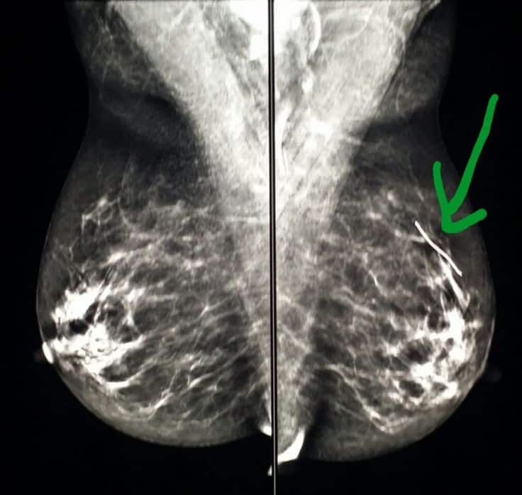
Many women experience a phone call from their breast imaging center. The call often concerns the patient coming back for additional imaging of tiny white spots called calcifications. Calcifications are frequently seen on mammograms they occur most often in women over 50. They may appear in any womans breasts and, occasionally, occur in a mans breast tissue.
Most breast calcifications are benign . However, a few patterns of calcification are suggestive of some precancerous conditions or, even, breast cancer.
You May Like: What Does Male Breast Cancer Lump Feel Like
Lets Do Some Q& A About Mammography
How are mammograms done?
During a mammogram, your breasts are compressed between two firm surfaces to spread out the breast tissue. Then, an x-ray captures black and white images of your breasts that are displayed on a computer screen and examined by a doctor who looks for signs of cancer.
How can mammograms be used?
A mammogram can be used either for screening or for diagnostic purposes.
How often should you have a mammogram?
It all depends on your age and your risk of breast cancer.
How do I know when I should begin screening mammography?
Some general guidelines for when to begin screening mammography include women with an average risk of breast cancer and woman with a high risk of breast cancer.
What are the risks?
Some known risks and limitations of mammograms include the following: Mammograms:-
- expose you to low-dose radiation
- are not always accurate
- can be difficult to interpret in younger women
- may lead to additional testing
- can not detect all breast cancers
- may show cancer, but not all of the tumors can be cured
How do I prepare for my mammogram appointment?
- Choose a certified mammogram facility
- schedule the test for a time when your breasts are least likely to be tender
- bring your prior mammogram images
- do not use deodorant before your mammogram
- consider an over-the-counter pain medication if you find that having a mammogram is uncomfortable.
What can a radiologist possibly find on my mammogram imaging?
Well, possible findings can include:-
- calcium deposits
Breast Fat Necrosis Vs Oil Cyst Symptoms

Oil cysts can also cause a lump in your breast and sometimes form along with fat necrosis.
Oil cysts are also noncancerous, fluid-filled sacs that form when the oils from decomposing fat cells collect in one place instead of hardening into scar tissue. Your body coats the oil sac with a layer of calcium , and the sac will feel:
Similar to a lump caused by fat necrosis, a lump is probably the only symptom youll notice with an oil cyst. These cysts might show up on mammograms, but theyre usually diagnosed with a breast ultrasound.
Oil cysts usually go away on their own, but your doctor can drain the fluid inside the cyst with a
of breast fat necrosis are of perimenopausal age and have pendulous breasts. Pendulous breasts have a longer shape and tend to droop downward more than other breast shapes.
Other demographic factors, such as race, are not associated with a higher risk of fat necrosis.
Fat necrosis is most common after breast surgery or radiation, so having breast cancer will raise your risk of fat necrosis. Breast reconstruction after cancer surgery may also increase your risk of fat necrosis.
Read Also: How Fast Breast Cancer Develops
The Following Statement Is Attributed To Binita Ashar Md Director Of The Office Of Surgical And Infection Control Devices In The Fdas Center For Devices And Radiological Health
- Statement From:
Today, the U.S. Food and Drug Administration issued a safety communication informing patients and providers about reports of squamous cell carcinoma and various lymphomas located in the capsule or scar tissue around breast implants. After an initial extensive review, we currently believe that the risk of SCC and other lymphomas occurring in the tissue around breast implants is rare. However, in this case, and when safety risks with medical devices are identified, we wanted to provide clear and understandable information to the public as quickly as possible.
In some reported cases, patients were diagnosed years after having breast implants and presented with findings such as swelling, pain, lumps or skin changes. These emerging reports of lymphoma in scar tissue are different from Breast Implant Associated Anaplastic Large Cell Lymphoma , which the FDA began communicating about as a potential risk more than a decade ago.
We know that breast implants are not lifetime devices, and that the longer a patient has breast implants, the more likely they will need to be removed or replaced. We also understand that information regarding breast implant risks can be overwhelming for a patient. For this reason, we encourage review of our website with attention to patient labeling, which has easy to understand information in the patient brochure.
Causes Of Breast Lumps That Arent Breast Cancer
For women, noticing a mass in the breast can be frightening, with your mind naturally leaping to breast cancer as the cause. But many breast masses arent related to breast cancer.
There are more than a dozen causes of breast masses that arent related to breast cancer, says Dean Currie, MD, general surgeon with West Tennessee Medical Group Jackson Surgical Associates. While any mass should be examined by a medical provider, its important to remember that there are many different types of benign breast masses. Its very common for a woman to notice a breast mass thats not cancerous.
But if a breast mass isnt related to cancer, what causes it? Lets take a deeper dive into the topic.
But First, Lets Touch on Breast CancerObviously, breast mass can be a sign of breast cancer. If you notice a breast mass during a self-exam or at another time, its important to let your OB/GYN or other medical provider know about it.
He or she will likely recommend you come in for a physical exam, and then depending on what is seen, you may also need to undergo a diagnostic mammogram, and often an ultrasound.
How can you tell if the breast mass you spot is cancerous? Well, you really cant. While noncancerous breast masses typically have smooth edges and can be moved manually with the fingers, that isnt always the case.
The only way to tell for certain is to have a provider take a look and order testing as necessary.
Here are a few common benign causes of breast masses:
Questions?
Recommended Reading: New Treatment For Her2-positive Breast Cancer
Fat Necrosis And Dystrophic Calcifications
Fat necrosis and dystrophic calcifications can be seen on the sonogram as a hypoechoic or hyperechoic irregular mass with acoustic shadowing. Correlation with mammographic images, surgical history, pathology findings, and clinical breast examination are important for accurate assessment. Serial ultrasounds and/or mammograms or biopsy may be needed.
Infectious Mastitis And Breast Abscess
Breast abscess is a complication of infectious mastitis. Abscesses can be associated with lactation, in the case of puerperal abscesses, or independent of pregnancy, in the case of nonpuerperal abscesses . Puerperal abscesses tend to be peripheral in location and are often easily recognized clinically. Nonpuerperal abscesses can pose a diagnostic challenge and are more commonly seen in younger women. They are usually periareolar and typically have worse outcomes and a higher rate of recurrence than puerperal abscesses. The risk factors for nonpuerperal breast abscesses are thought to include smoking and diabetes .
Mammographically, mastitis and breast abscess present with skin thickening, asymmetry, a mass, or architectural distortion . Sonographic features of breast abscesses include one or more hypoechoic collections of variable shapes and sizes that are often continuous and multiloculated . Breast abscesses typically demonstrate a thick echogenic rim and increased vascularity, suggesting malignancy . Associated mastitis presents as an area of increased parenchymal echogenicity, representing inflamed glandular parenchyma. Skin thickening, distended lymphatic vessels, and inflammatory axillary adenopathy can also be seen. On MRI, breast abscesses will typically be T2-hyperintense, have progressive enhancement kinetics, and sometimes have the characteristic thin rim of peripheral enhancement .
Fig. 2
You May Like: Does Breast Cancer Have Any Symptoms