Getting A Second Opinion
No one knows your body better than you. If your biopsy results are malignant, or even if theyre benign, its always fine to get a second opinion, and is usually a good idea.
Make sure to see a specialist. You can take your mammogram results to a breast imaging center to be reexamined by a breast imaging radiologist or see another doctor. Ask your insurance how this will be covered.
Your doctor may even recommend you get a second opinion, especially if you have had cancer or have a family history of cancer.
I Had A Breast Biopsy And Was Told That The Calcium Deposit Were Suspicious What Does That Mean
Ask U.S. doctors your own question and get educational, text answers â it’s anonymous and free!
Ask U.S. doctors your own question and get educational, text answers â it’s anonymous and free!
HealthTap doctors are based in the U.S., board certified, and available by text or video.
A Warning Sign Of Early Breast Cancer
In some cases, calcifications on a mammogram represent the earliest form of breast cancer, which is called ductal carcinoma in situ . In DCIS, the cancerous cells are confined to the milk ducts of the breast. DCIS is very treatable and highly curable but in some cases, if left untreated, it has the potential to become invasive breast cancer.
During a mammogram, radiologists will also look for masses or other changes in the tissue that have developed since your last screening, and they will make an assessment of your breast density. Breasts are considered dense if the glandular and fibrous tissue makes up more than 50 percent of the breast tissue. Patients with extremely dense breasts may be at increased risk of breast cancer.
Tari A. King, MD, FACS, is Chief of Breast Surgery at Dana-Farber/Brigham and Womens Cancer Center, Associate Division Chief for Breast Surgery and Associate Chair for Multidisciplinary Oncology at Brigham and Womens Hospital, and Associate Professor of Surgery at Harvard Medical School.
Also Check: Breast Cancer Metastasis To Lymph Nodes Prognosis
Breast Cancer Calcifications: Identification Using A Novel Segmentation Approach
Melkamu Teshome Ayana
1University of Engineering and Management, Kolkata, India
2School of Computer Science and Engineering, Lovely Professional University, Phagwara, Punjab, India
3Mehran University of Science and Technology, Jamshoro, Pakistan
4University of Engineering and Management, Kolkata, India
5Department of Hydraulic and Water Resources Engineering, Arba Minch University, Ethiopia
Abstract
1. Introduction
AI and machine learning are recently widely used in health care for the prediction of critical diseases like colorectal cancer, Alzheimer, fetal brain abnormality detection, and type-2 diabetes risk prediction, and the present study used AI and ML for breast cancer prediction.
Breast cancer was expected to claim an estimated 40,870 lives in 2005 . Breast cancer is the second leading cause of mortality from cancer in women. According to the most current statistics, death rates decreased substantially between 1992 and 1998, with the greatest reductions occurring among younger white and black women.
1.1. Automated Breast Ultrasound Technique
The existing techniques for breast cancer detection, such as mammograms, ultrasounds, and biopsies, were time-consuming, necessitating the development of a computerized diagnostic system using machine learning technology. This approach incorporates algorithms that aid in tumor categorization and cell detection more precisely and efficiently.
1.2. Breast-Specific Gamma Imaging
1.3. Scintimammography
1.4. Sonography
Vacuum Assisted Excision Biopsy

Occasionally, you may have a vacuum assisted excision biopsy to remove an area of calcification. This is a similar procedure to a vacuum assisted biopsy, but more tissue may be removed.
This may mean that an operation under a general anaesthetic can be avoided.
Don’t Miss: Signs Of Breast Cancer Recurrence After Mastectomy
How Are High Calcium Levels Diagnosed And How Are They Managed
Your doctor can do a blood test to learn if you have a high calcium level. You may also have blood tests to check how well your kidneys are working. Your doctor will treat a high calcium level if you have it. The treatment depends on how severe the condition is.
Mild hypercalcemia
People who have no symptoms receive extra fluids, usually given through a vein. This will help your kidneys remove extra calcium from your blood.
Moderate or severe hypercalcemia
This can be treated by:
-
Continuing cancer treatment.
-
Replacing fluids lost through vomiting and urination.
-
Taking medication to stop bone from breaking down. You may be prescribed a bisphosphonate, such as zoledronic acid , pamidronate , or ibandronate , or denosumab . Talk with your doctor about the risks and benefits of taking such medications.
-
Taking medicine called steroids. These can help stop bone from breaking down. They also help your bones take more calcium from your food. Steroids can also raise your risk of bone loss over time. Talk with your doctor about the risks and benefits of taking steroids.
-
Taking a hormone called calcitonin. This hormone functions by reducing calcium release from your bones and increase calcium secretion from your kidneys.
-
Using dialysis if you haves kidney failure. Dialysis is a machine-based process that cleans your blood when your kidneys are not working properly.
The Operation Removing The Calcification
Once the hookwire is in position you will be taken to the operating theatre. The operation will be performed under general anaesthetic. The surgeon will remove the piece of tissue at the tip of the wire. Once the tissue is removed, it will be returned to the imaging department where an X-ray of the tissue will be performed to confirm that the calcification is in the tissue that has been successfully removed. The specimen will then be sent to the pathologist.
Post hookwire insertion
You will need the Adobe Reader to view and print these documents.
-
Meet
Recommended Reading: Bob Red Mill Baking Soda Cancer
What Happens If My Doctor Finds Breast Calcifications On My Mammogram
If you have macrocalcifications, no further testing or treatment is needed, because they are not harmful. If microcalcifications are seen on your mammogram, another mammogram may be performed to get a more detailed look at the area in question. The calcifications will be determined to be either “benign,” “probably benign,” or “suspicious.”
About Calcium In Your Body
Everybody needs calcium for many body functions. It helps form bones and teeth, and it also helps your muscles, nerves, and brain work correctly. Most of the calcium in your body is in your bones. Normally, your blood contains only a small amount. When you are healthy, your body controls the level of calcium in your blood.
Cancer can cause a high calcium level in the blood in several ways. High calcium levels due to cancer are not caused by too much calcium in your diet. Eating fewer dairy products and other high-calcium foods will not lower high blood calcium levels.
Cancers that more commonly cause high calcium levels in your blood include:
-
Lung cancer
-
Loss of consciousness
-
Coma
You and your family should know these serious symptoms. Ask your doctor what you should watch for and when to get treatment.
Read Also: Is Estrogen Positive Breast Cancer Hereditary
What Does It Mean To Have Scattered Fibroglandular Tissue
Medically reviewed by Yamini Ranchod, PhD, MS on February 27, 2019 Written by Kimberly Holland. Scattered fibroglandular tissue refers to the density and composition of your breasts. A woman with scattered fibroglandular breast tissue has breasts made up mostly of non-dense tissue with some areas of dense tissue.
Should I Be Concerned
There are two main types of calcifications:
- Macro: Macrocalcifications appear large and round on a mammogram. Usually theyre not related to cancer at all.
- Micro: Microcalcifications are small and may appear in clusters. They are usually benign, but could be a sign of breast cancer.
We see breast calcifications all the time. Most of the time they are harmless and arent cause for concern.
Don’t Miss: How Treatable Is Breast Cancer
What Happens During A Breast Biopsy
Two types of biopsies are used to remove breast calcification tissue for further study, including stereotactic core needle biopsy and surgical biopsy.
Core needle biopsy: Under local anesthesia a radiologist, using a thin, hollow needle and guided by a computer imaging device, will remove a small piece of tissue containing the suspicious calcifications.
Surgical biopsy: If tissue cannot be successfully removed using a core needle biopsy or the results are unclear, surgery may be needed to get a sample of the calcified breast tissue. A surgeon will perform the biopsy in an operating room under local or general anesthesia. Prior to the surgical procedure, a radiologist may use X-rays to identify the calcified breast tissue and will then mark the tissue to be removed — with either a thin wire or with dye. A surgeon will then cut the tissue sample so that it can be sent to a lab for analysis.
If you have breast calcifications, talk to your doctor about your concerns.
Show Sources
Before The Operation Inserting The Hookwire

The hookwire will be inserted by a radiologist. Before the operation, you will go to the breast department. The abnormal area in the breast will be identified with a mammogram. An injection of local anaesthetic will be given to numb part of the breast and the hookwire will be inserted under the guidance of the mammogram. A mammogram will also be performed after the wire has been inserted. This mammogram is performed with minimal pressure on the breast so that it does not make the wire move. This checks the position of the wire and helps the surgeon plan the operation. Once the wire is in position, it will be taped in place and a dressing will be applied.
Hookwire inside insertion needle
Don’t Miss: Average Life Expectancy Stage 4 Breast Cancer
What Causes Breast Calcifications
It is not known what causes calcifications to develop in breast tissue, but they are not caused by eating too much calcium or taking too many calcium supplements.
They are seen on mammograms of about half of all women over age 50. However, they also are seen in about 10 percent of mammograms on younger women. Women who have had breast surgery for any reason or who have injured their breasts, such as in a car accident, seem to be at higher risk for developing calcifications, as are women who have been treated for breast cancer in the past. Calcifications may also occur within vessels in the breast related to older age or from a past infection in the breast tissue.
How Are Breast Calcifications Detected
Calcifications are a common find on a mammogram, with increasing prevalence after the age of 50. There are a variety of causes for calcifications, including:
- Aging
- Infection
- Inflammation
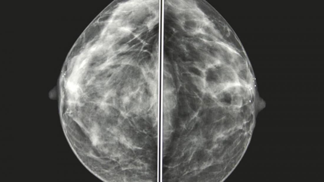
Calcifications, unlike lumps, cannot be detected using touch. They can only be found using mammography or, rarely, ultrasounds.
Also Check: Chemotherapy For Stage 2 Breast Cancer
Questions About Breast Calcifications Answered
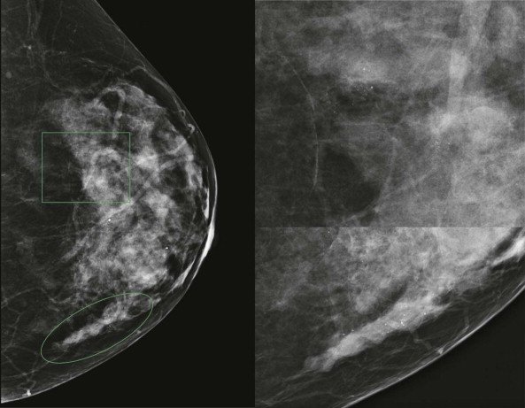
Breast calcifications are calcium deposits found through screening mammograms. When calcium builds up in soft tissue, it can appear like small white specks or salt crystals on diagnostic images. These spots can be found in various organs, such as the lungs or brain, but theyre commonly found in breast tissue with screening mammograms.
Breast calcifications are pretty common, but most people dont know they have them unless they have been mentioned on prior mammogram reports, says
So, are these white spots a sign of cancer? Here, Dryden answers this and three more questions about breast calcifications.
Are breast calcifications a sign of cancer?
Theyre often benign, but calcifications can sometimes be an earlysign of breast cancer. The most common form of cancer we see with calcifications is ductal carcinoma in situ, which is considered stage 0 cancer, Dryden says.
Benign calcifications are often scattered throughout both breasts. If one breast has calcifications and the other doesnt, that could be a sign that we need to take a closer look at them. Breasts are often symmetrical, so when we see that one breast has calcifications and the other doesnt, that could be a red flag, Dryden says.
Calcifications can also be a sign of non-cancerous conditions and may represent a benign process. Fibrocystic breasts, which feel lumpy or rope-like in texture, can also be associated with calcifications.
What causes breast calcifications?
Can I prevent breast calcifications?
Do Breast Calcifications Mean That I Have Breast Cancer
Categories:Breast Cancer,Cancer Screening
Many women experience a phone call from their breast imaging center. The call often concerns the patient coming back for additional imaging of tiny white spots called calcifications. Calcifications are frequently seen on mammograms they occur most often in women over 50. They may appear in any woman’s breasts and, occasionally, occur in a man’s breast tissue.
Most breast calcifications are benign . However, a few patterns of calcification are suggestive of some precancerous conditions or, even, breast cancer.
Don’t Miss: Can Stage 3 Breast Cancer Be Cured
How Are Breast Calcifications Treated
If the calcifications look benign, nothing more needs to be done. They dont need to be removed and wont cause you any harm.
If the calcifications look indeterminate or suspicious you will need further tests, as in many cases a mammogram wont give enough information. This doesnt mean something is wrong, but further tests will help to make an accurate diagnosis.
These tests are usually done in the breast clinic or x-ray department as an outpatient. You wont have to stay overnight in hospital.
Further tests could include the following:
Do Breast Calcifications Increase My Risk Of Breast Cancer
Most breast calcifications are due to benign changes, which does not increase your risk of breast cancer.
However, if the breast calcifications are due to atypical change, this may slightly increase your risk of breast cancer.
Your treatment team will be able to discuss this further with you.
Its important to continue to be breast aware, and to go back to your GP if you notice any changes in your breasts, however soon this is after you were told you had calcifications.
Recommended Reading: What Organs Does Breast Cancer Affect
Calcification Of My Left Breast
Good Morning
I went to Grimsby hospital breast clinic on the 23rd January just for a check up how little did I know it would change my life!!
At 11:15 am I was told after my mammogram I had calcification on my left breast and this was cancer! once you hear that word your World explodes inwards, it did for me, the next 1 hour was shell shock, I had my boyfriend with me so he was able to ask questions and be there to give me support, I was told I needed an ultrasound which lead to me having 3 needle biopsies.
After seeing the consultant for the second time I was informed I would have to go back next Wednesday for the results and options but when asked about what he recommended it was a full mastectomy.
The most scary part is although the nurses were there to offer comfort at no time did anyone tell me where I could go to get help? it is fine having all these leaflets in the waiting room but to be honest the last thing on my mind was money or other things, I just wanted someone to explain in simple words exactly what i had? yet no one did this I was just told you have cancer now go home and come back next week?
Am i wrong here? do i expect to much from our goverment and NHS? I just go home and what carry on as if nothing as happened and thing well it is just like having a cold it will go away soon?
So please can anyone help me with what is it I have? and what do I do now?
Regards
Hi Elaine
Best wishes Ann
Hello Elaine and welcome.
Annabel.
Arterial Calcifications And Heart Disease

Calcifications believed to be in the arteries of the breast have traditionally been thought of as incidental findings not associated with breast cancer risk, so they didn’t get much attention. However, that’s changing.
Research from 2014 suggests that the presence of breast arterial calcifications is associated with underlying coronary artery disease in women over 40 who don’t have any symptoms of heart disease. Their presence was even more likely to predict the presence of arteriosclerosis than risk factors such as high blood pressure, a family history of heart disease, and more.
Unfortunately, symptoms of coronary artery disease or a heart attack in women are often different from what is considered “typical,” and symptoms such as profound fatigue, nausea, or even jaw pain may be the only ones heralding these concerns. Mammograms may, by finding arterial calcifications, help in detecting coronary artery disease before problems occur.
Since much of the research looking at the meaning of breast arterial calcifications is relatively new, it’s important to be your own advocate and ask questions if you should see a note about these on your report.
You May Like: Stage Zero Breast Cancer Survival Rate
Why Do Breast Calcifications Form
Calcifications are usually non-cancerous changes in the breast tissue associated with aging. But there are various causes for calcifications your breast health provider will work to determine the cause of any breast calcification to ensure that the changes seen on a patient’s mammogram are not cancerous.
What Are The Features Of Scattered Breast Tissue
Features of scattered fibroglandular breast tissue may include: 1 lumps in the breasts 2 cysts, which are fluid-filled round or oval sacs 3 fibrosis, or prominent scar-like fibrous tissue 4 an overgrowth of cells in the lining of the milk ducts or milk-producing tissues 5 enlarged breast lobules, or adenosis More
Read Also: Can Nipple Piercing Cause Cancer
Breast Changes Of Concern
Some breast changes can be felt by a woman or her health care provider, but most can be detected only during an imaging procedure such as a mammogram, MRI, or ultrasound. Whether a breast change was found by your doctor or you noticed a change, its important to follow up with your doctor to have the change checked and properly diagnosed.
Check with your health care provider if your breast looks or feels different, or if you notice one of these symptoms:
- Lump or firm feeling in your breast or under your arm. Lumps come in different shapes and sizes. Normal breast tissue can sometimes feel lumpy. Doing breast self-exams can help you learn how your breasts normally feel and make it easier to notice and find any changes, but breast self-exams are not a substitute for mammograms.
- Nipple changes or discharge. Nipple discharge may be different colors or textures. It can be caused by birth control pills, some medicines, and infections. But because it can also be a sign of cancer, it should always be checked.
- Skin that is itchy, red, scaled, dimpled or puckered