How Do You Fix Breast Asymmetry Without Surgery
If youre bothered by overly large, heavy breasts, a breast reduction can help reduce overall size and minimize disparity between two sides. If this is not a concern, fat grafting may be suitable. This technique uses your own natural fatty tissue from another area of the body and transfers it into the breasts.
How Is Breast Density Categorized
Doctors use the Breast Imaging Reporting and Data System, called BI-RADS, to group different types of breast density. This system, developed by the American College of Radiology, helps doctors to interpret and report back mammogram findings. Doctors who review mammograms are called radiologists. BI-RADS classifies breast density into four categories, as follows:
- Almost entirely fatty breast tissue, found in about 10% of women
- Scattered areas of dense glandular tissue and fibrous connective tissue found in about 40% of women
- Heterogeneously dense breast tissue with many areas of glandular tissue and fibrous connective tissue, found in about 40% of women
- Extremely dense breast tissue, found in about 10% of women
If you are told that you have dense breasts, it means that you have either heterogeneously dense or extremely dense breasts.
The four breast density categories are shown in this image. Breasts can be almost entirely fatty , have scattered areas of dense fibroglandular breast tissue , have many areas of glandular and connective tissue , or be extremely dense . Breasts are classified as âdenseâ if they fall in the heterogeneously dense or extremely dense categories.
What Percentage Of Mammogram Callbacks Are Cancer
Of all women who receive regular mammograms, about 10 percent will get called back for further testing and of those, only about 0.5 percent will be found to have cancer. Your chances of being diagnosed with cancer after a callback are small, but your doctor wants to be sure there is no cancer present.
Read Also: What Is The Rarest Form Of Breast Cancer
Should Women With Dense Breasts Have Additional Screening For Breast Cancer
In some states, mammography providers are required to inform women who have a mammogram about breast density in general or about whether they have dense breasts. Many states now require that women with dense breasts be covered by insurance for supplemental imaging tests. A United States map showing information about specific state legislation is available from DenseBreast-info.org.
Nevertheless, the value of supplemental, or additional, screening tests such as ultrasound or MRI for women with dense breasts is not yet clear, according to the Final Recommendation Statement on Breast Cancer Screening by the United States Preventive Services Task Force. Ongoing clinical trials are evaluating the role of supplemental imaging tests in women with dense breasts. NCIâs Cancer Information Service can tell you about clinical trials and provide tailored clinical trial searches to help you learn more about clinical trials related to breast density and breast cancer screening.
Recent research has suggested that for women with dense breasts, a screening strategy that also takes into account a womanâs risk factors and protective factors may be the best predictor of whether a woman will develop breast cancer after a normal mammogram and before her next scheduled mammogram.
As you talk with your doctor about your personal risk for breast cancer, keep in mind that:
- risk factors increase your chance of breast cancer
- protective factors lower your chance of breast cancer
When To See A Doctor

Most changes to the size of your breasts are caused by changes in your hormones, which will naturally correct on their own.
However, if these changes in your uneven boobs do not go away or if you experience any of the following symptoms, you should visit your doctor as they will be able to check for any possible health problems:
- Swelling in one of your breasts
- Pain in your chest area
- Nipple discharge or fluid
- Abnormal changes to the nipple
- A one-sided lump on one of your breasts that has suddenly appeared
- The variation between the size of your breasts is significantly large
- The skin around the breast is red, itchy, flaky or firm and dimpled
- The tissue around the breast or under the arm feels firm or thick
Speak to a patient advisor
Don’t Miss: Can Sleeping With Your Bra On Cause Breast Cancer
Breastfeeding And Uneven Breast Size
Experiencing a certain amount of breast asymmetry is a normal part of breast feeding, especially if one of your breasts receives more stimulation than the other as a result of your baby preferring one breast over the other or if you feed on the same breast most of the time.
This can cause one breast to grow larger as it produces more milk, and alternating which breasts you feed from can help to avoid this problem.
For most breastfeeding mothers, asymmetrical breasts are not a medical concern. However, if one of your breasts has remained smaller from the beginning of your pregnancy and did not get any larger, visit your doctor for a consultation.
If You Have An Abnormal Cbe:
If youre under 30 and your CBE reveals a lump, your doctor may recommend starting with observation. This means waiting 1-2 menstrual periods to see if the lump goes away on its own, then getting checked again. If youre not comfortable waiting, let your doctor know or ask for a second opinion.
If youre 30 or older and your CBE reveals a lump or other change, the most likely next step is a follow-up mammogram and maybe a breast ultrasound.
Recommended Reading: What Is The Last Stage Of Breast Cancer
How Often Is Breast Asymmetry Cancer
Asked by:Haven Cummings
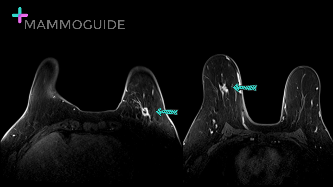
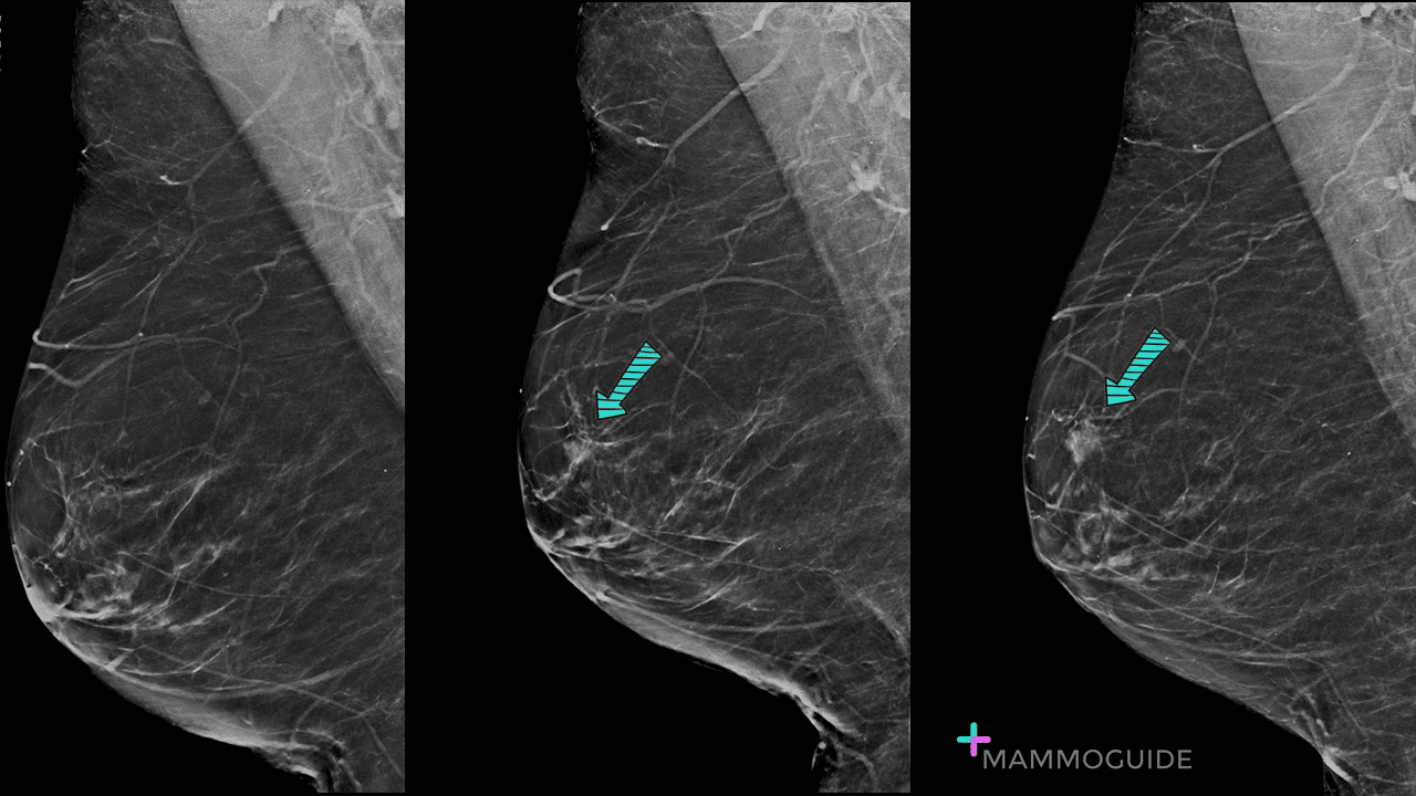
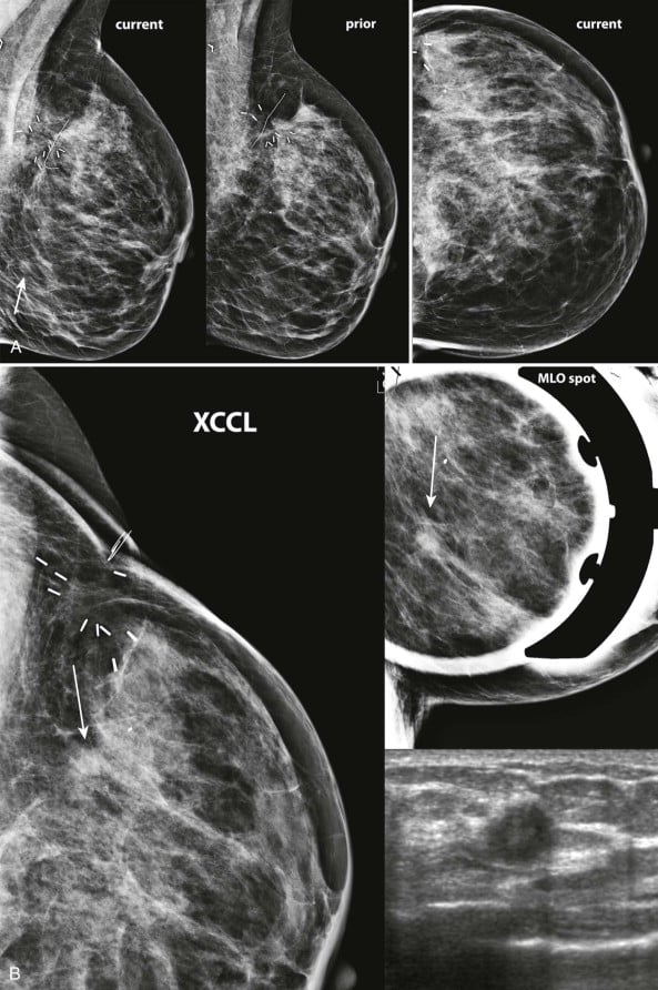
In mammography, an asymmetry is an area of increased density in 1 breast when compared to the corresponding area in the opposite breast. Most asymmetries are benign or caused by summation artifacts because of typical breast tissue superimposition during mammography, but an asymmetry can indicate breast cancer.
Causes Of Breast Asymmetry
During puberty, the left and right breast often develop at a slightly different pace. Breasts may appear asymmetrical until they have finished growing, or they may remain different shapes and sizes throughout a persons life.
Hormonal changes can cause one or both breasts to change at any point in a persons life, for example:
- at specific points in the menstrual cycle
- during or near menopause
- during pregnancy or breast-feeding
- when using a hormonal contraceptive, such as birth control pills
Breasts that change size or shape because of hormones often return to normal. Hormonal changes can also cause breasts to feel lumpy or lose fat and tissue. However, if these changes do not go away, it is a good idea to visit the doctor to who will check for any possible health problems.
Some underlying conditions that can affect breast size and shape include:
- Tubular breasts: Also called breast hypoplasia, tubular breasts can develop in one or both breasts during puberty.
- Amastia or amazia: A condition that causes problems in the development of breast tissue, the areola, or nipple.
- Poland Syndrome: Where a chest muscle does not develop properly, which can affect the breast on one side of the body.
Read Also: Can Binding Cause Breast Cancer
What Is Breast Asymmetry
Literally speaking, âbreast asymmetryâ means that the two breasts are not mirror-image-identical to one another but differ in one way or another. In truth, this is actually completely normal. In fact, the vast majority of women have uneven breasts. The problem occurs when that unevenness is so marked that it becomes very obvious.
Breast asymmetry occurs when a woman has one breast that is a different size, position or volume to the other. It is a very common condition that affects more than half of all women. There are three types of breast asymmetry:
Understanding Your Mammogram Report
Lisa Jacobs, M.D., Johns Hopkins breast cancer surgeon, and Eniola Oluyemi, M.D., Johns Hopkins Community Breast Imaging radiologist, receive many questions about how to interpret common findings on a mammogram report. The intent of the report is a communication between the doctor who interprets your mammogram and your primary care doctor. However, this report is often available to you, and you may want to better understand it. Both experts suggest that you sit down with your doctor to discuss the findings of the report to avoid confusion.
Here are answers to 10 of the most commonly asked questions:
Read Also: Does Breast Cancer Cause Arm Pain
Putting Your Mind At Ease
Many women feel anxious and uncertain while theyâre getting follow-up exams and waiting for test results.
Doctors say that learning about the tests and writing down questions to bring to your appointments can help you feel calmer and more in control. They also recommend asking someone you trust to come with you, as a second set of ears when you talk with your doctor. That person can also take notes for you and offer their support.
Show Sources
What Are Clip Markers And Why Are They Used During Biopsies

After a mammogram screening, a small percentage of women will have afinding that may require additional diagnostic imaging. This is called arecall. If a patient is recalled, additional imaging will be performed, andonly about 2 percent of women may need a biopsy. During a biopsy, aradiologist with breast imaging expertise inserts a small metallic clip inthe breast to help locate the biopsy site in case further testing isneeded.
Also Check: Can Breast Cancer Cause Arm Numbness
Combined Digital Mammography And 3d Tomosynthesis Findings
Combined digital mammography and 3D tomosynthesis BIRADS category was given for each lesion according to the BIRADS mammography morphology descriptors 37/57 lesions were considered benign , while 20/57 lesions were considered malignant.
After revising the pathology results 15/18 lesions were true positives, 5/39 lesions were false positive, 3/18 lesion was false negative, and 34/39 lesions were true negatives.
The false-positive results are less when compared to digital mammography alone as tomosynthesis overcame the tissue overlap in focal asymmetries.
So, combined digital breast mammography and 3D tomosynthesis had a sensitivity of 88.33%, a specificity of 87.18%, a positive predictive value of 75.00%, and a negative predictive value of 91.89%.
Many researchers have investigated the potential role of DBT in both screening and diagnostic settings. Improvements in sensitivity and specificity are expected after adding DBT to conventional mammography because DBT eliminates overlapping tissues, and lesion margins can be more readily assessed, which may reduce the need for extra views as results of Kim et al .
Is It Common To Be Called Back For An Ultrasound After A Mammogram
Getting called back after a screening mammogram is fairly common, and it doesnt mean you have breast cancer. In fact, fewer than 1 in 10 women called back for more tests are found to have cancer. Often, it just means more x-rays or an ultrasound needs to be done to get a closer look at an area of concern.
Also Check: What Color Is Breast Cancer Pink
Technique Of 3d Tomosynthesis
For 3D digital tomosynthesis, two views were obtained. 3D DBT involved the acquisition of 12 to 15 2D projection exposures by a digital detector from a mammographic x-ray source which moves over a limited arc angle. The 3D volume of compressed breast was reconstructed from the 2D projections in the form of series of images through the entire breast. Images were assessed in the workstation.
Image Analysis And Interpretation Of Breast Ultrasound
Each lesion was evaluated regarding shape, boundary, margin, echo pattern, orientation and posterior acoustic features, calcifications, and axillary lymph nodes. The BIRADS category of each lesion was determined according to BIRADS atlas of Ultrasound 2013, guided by the results of clinical data and breast ultrasound findings but blinded to the final pathological diagnosis.
Reference points for us were histopathological analysis of biopsy and surgical samples, fine-needle aspiration cytology, or close follow-up.
Also Check: Does Breast Cancer Cause Heartburn
What Does Asymmetry In Left Breast Mean
Breast asymmetry occurs when one breast has a different size, volume, position, or form from the other. Breast asymmetry is very common and affects more than half of all women. There are a number of reasons why a womans breasts can change in size or volume, including trauma, puberty, and hormonal changes.
What Should I Do If I Notice Abnormal Changes Or Symptoms Even After Mymammogram Comes Back Normal
Breast self-exams are important because they allow you to get to know yourbreasts and their normal appearance. If you notice abnormal symptoms orchanges to your breast geography, request additional testing. Do not ignoreabnormal breast changes or symptoms, such as discharge or a lump, but keepin my mind that several lifestyle changes, such as weight gain, weightloss, hormone changes and hormone replacement therapy, can cause yourbreasts to change.
Note: The radiologist may call you back after a baseline mammogram for additional testing because he or she hasnothing to compare the mammogram to. This will also help identify changesto your breasts over time.
Also Check: Is Male Breast Cancer Hereditary
How Common Are Dense Breasts
Nearly half of all women age 40 and older who get mammograms are found to have dense breasts. Breast density is often inherited, but other factors can influence it. Factors associated with lower breast density include increasing age, having children, and using tamoxifen. Factors associated with higher breast density include using postmenopausal hormone replacement therapy and having a low body mass index.
Terminology And Significance Of Types

There are four types of asymmetries: global asymmetry, focal asymmetry, developing asymmetry, and one-view asymmetry . Differentiation between the types is important because the positive predictive value for malignancy and management differs according to type. Asymmetry may be global or focal . If an asymmetry is new compared with old mammograms, it is considered a developing asymmetry. If an asymmetry is only seen on a single projection, it is a one-view asymmetry.
-
One-view asymmetry
Asymmetries do not fulfill the criteria for the other soft tissue density findings described in the BI-RADS Atlas. They lack the convex borders of masses and are often interspersed with fat . They also lack the radiating lines or tissue retraction of architectural distortion and the tubular branching appearance of a dilated duct.
Don’t Miss: What Is The Medical Term For Breast Cancer
High Prolactin Hormone Level
Hyperprolactinemia means the pituitary gland secretes too much prolactin, the hormone responsible for producing milk in a new mother. The condition can appear in both women and men.
It can be caused by pregnancy by an ovulatory disorder by some psychiatric medications or by a prolactin-secreting tumor of the pituitary
Women with other reproductive disorders, such as polycystic ovary syndrome are most susceptible. Hyperprolactinemia is also seen in those with hypothyroidism and chronic renal failure. Many patients on hemodialysis have elevated prolactin levels.
Symptoms in both women and men include reduced libido and infertility. Men may show breast enlargement and women may develop breast milk.
If not treated, hyperprolactinemia can result in loss of bone density in both women and men.
Diagnosis is made through blood testing to measure hormone levels, and sometimes MRI of the pituitary gland underneath the brain.
Treatment may include “watchful waiting,” or a period spent observing the symptoms to see if they change drug therapy or surgery.
Combined Digital Mammography 3d Tomosynthesis And Ultrasound Findings
Combined digital mammography, 3D tomosynthesis, and ultrasound BIRADS category was given for each lesion according to the BIRADS mammography morphology descriptors 36/57 lesions were considered benign while 21/57 lesions were considered malignant.
After revising the pathology results, 18 lesions were true positives, 3 lesions were false positive, 0 lesions was false negative, and 36 lesions were true negatives.
Kim et al. found that previous prospective clinical studies have demonstrated that appropriate use of US as an adjunct to mammography improves sensitivity and specificity of breast cancer diagnoses, particularly in women with dense breasts and in younger women.
In this study, combined digital mammography, 3D tomosynthesis, and ultrasound had a sensitivity of 100.00%, a specificity of 92.31%, a positive predictive value of 85.71%, and a negative predictive value of 100.00%.
However, some points made were usage of 3D is limited, like relatively higher dose of radiation, higher cost, and less availability than FFDM. Considering this study, decreased number of patients may make the results a matter of discussion.
Read Also: Does Underwire Cause Breast Cancer
What Gets Stored In A Cookie
This site stores nothing other than an automatically generated session ID in the cookie no other information is captured.
In general, only the information that you provide, or the choices you make while visiting a web site, can be stored in a cookie. For example, the site cannot determine your email name unless you choose to type it. Allowing a website to create a cookie does not give that or any other site access to the rest of your computer, and only the site that created the cookie can read it.
What Will Happen At The Follow
- Youll likely get another mammogram called a diagnostic mammogram. A diagnostic mammogram is done just like a screening mammogram, but more pictures are taken so that any areas of concern can be looked at more closely. A doctor called a radiologist will be on hand to advise the technologist , to be sure they have all the images that are needed.
- You may also get another imaging test, such as an ultrasound of the breast, which uses sound waves to make pictures of the inside of your breast at the area of concern.
You will most likely be given the results of your tests during the visit. You might be told one of the following:
- The suspicious area on the mammogram turned out to be nothing to worry about, and you can return to your normal mammogram schedule.
- The area is probably nothing to worry about, but you should have your next imaging test sooner than normal usually in about 6 months to watch the area closely and make sure it’s not changing over time.
- The area could be cancer, so you will need a biopsy to know for sure.
Youll also get a letter with a summary of the findings that will tell you if you need more tests and/or when you should schedule your next mammogram.
Don’t Miss: Does Nipple Pinching Cause Breast Cancer