What Are Some Common Uses Of The Procedure
- Determining the Nature of a Breast AbnormalityDoctors use breast ultrasound to help diagnose breast abnormalities detected during a physical exam. These may include a lump or spontaneous bloody/clear nipple discharge. They also use ultrasound to characterize potential abnormalities seen on mammography or breast magnetic resonance imaging .Breast ultrasound can help determine if an abnormality is solid , fluid-filled , or both cystic and solid.
Breast ultrasound can be offered as a screening tool for women who:
- are at high risk for breast cancer and unable to undergo an MRI exam.
- are pregnant or should not be exposed to x-rays .
- have increased breast density when the breasts have a lot of glandular and connective tissue and not much fatty tissue .
- Ultrasound-guided Breast Biopsy When a breast ultrasound reveals a suspicious abnormality, the radiologist may recommend an ultrasound-guided biopsy. Because ultrasound provides real-time images, doctors often use it to guide biopsy procedures. A breast ultrasound exam will usually be necessary before the biopsy to plan the procedure and to determine if this method can be used.
After Your Breast Ultrasound
You can get dressed straight after the ultrasound. A specialist looks at the ultrasound pictures.
You might not need any further tests if everything looks normal. If the test shows a fluid-filled lump, a doctor or nurse might drain the fluid with a needle.
If a solid lump shows on the scan you might need to have more tests. These might include a breast x-ray or taking a sample of cells from the abnormal area . If the lymph nodes in your armpit look abnormal, you might have a needle biopsy in this area.
In a one-stop clinic you have these tests during the same visit.
Your doctor may suggest you have other tests or scans, for example an MRI. These are usually booked for another day.
At What Point Are You Considered Cancer Free
In a complete remission, all signs and symptoms of cancer have disappeared. If you remain in complete remission for 5 years or more, some doctors may say that you are cured. Still, some cancer cells can remain in your body for many years after treatment. These cells may cause the cancer to come back one day.
Don’t Miss: Does Nipple Pinching Cause Breast Cancer
Note To Users Outside Of The United States
WebMD and our technical infrastructure are located in the United States. The personal information that you provide to us is stored on servers located in the United States. If you are located in another jurisdiction, you should be aware that in order to provide the Services to you, we must transfer your personal information to the United States where it will be stored and processed in accordance with this Privacy Policy. We may transfer your information outside the United States to service providers with operations in other countries. By using the Services, you consent to such collection, storage and processing in the United States and elsewhere, though the United States and other jurisdictions may not afford the same level of data protection as considered adequate in your own country. We will take reasonable steps to protect your personal information. Note that your personal information may be available to the United States government or its agencies under legal process made in the United States.
Screening And Detection Rates Using Ultrasound

The discovery rate of malignant cancer using breast cancer screening methods is actually very low.
So, for example, the rate of detecting breast cancer on a mammogram is about 5 cancers in every thousand women screened.
Ultrasound alone for breast cancer screening detects slightly fewer malignancies. This tends to suggest that mammography is slightly more reliable in the detection of breast cancer.
Unfortunately, I have to stress here that the combination of mammography, ultrasound and even MRI can not completely exclude the possibility of breast cancer.
Indeed, up to 3% of women with suspicious lesions but negative mammograms and ultrasounds may still have breast cancer.
Read Also: Breast Cancer In Other Breast After Mastectomy
Breast Ultrasound To Screen For Breast Cancer
The doctors of The Tyanna OBrien Center for Womens Imaging at Mercy in Baltimore, Maryland offer screening and diagnostic breast ultrasound. Our doctors work collaboratively with the breast surgeons of The Hoffberger Breast Center at Mercy to provide comprehensive care for women.
Diagnostic breast ultrasound is used to evaluate breast symptoms or to evaluate abnormal findings on mammography. Ultrasound also is utilized for breast cancer screening in selected patients.
Stage Of Breast Cancer
When breast cancer is diagnosed, your doctors will give it a stage. The stage describes the size of the cancer and how far it has spread, and is used to predict the outlook.
Ductal carcinoma in situ is sometimes described as stage 0. Other stages of breast cancer describe invasive breast cancer and include:
- stage 1 the tumour measures less than 2cm and the lymph nodes in the armpit are not affected. There are no signs that the cancer has spread elsewhere in the body
- stage 2 the tumour measures 2 to 5cm, the lymph nodes in the armpit are affected, or both. There are no signs that the cancer has spread elsewhere in the body
- stage 3 the tumour measures 2 to 5cm and may be attached to structures in the breast, such as skin or surrounding tissues, and the lymph nodes in the armpit are affected. There are no signs that the cancer has spread elsewhere in the body
- stage 4 the tumour is of any size and the cancer has spread to other parts of the body
This is a simplified guide. Each stage is divided into further categories: A, B and C. If you’re not sure what stage you have, talk to your doctor.
Also Check: When Should You Start Screening For Breast Cancer
Dump Your Diy System For The Qa Tool Of Your Dreams
Go from hot mess to no stress with built-in QA and credentialing tools to automatically track and manage your POCUS program. With Exo Works, ultrasound exams are instantly available for review and collaboration wherever you arework or home. Fast loading images, simple markup tools and image tagging makes QA review a breeze and real-time feedback a reality.
Combining Ultrasound For Breast Cancer With Mri Or Biopsy
Research shows that the combination of ultrasound with Magnetic Resonance Imaging is a particularly good combination in follow-up evaluation of lesions found on mammography.
The detail of the MRI greatly assists in diagnostic and treatment decisions. Ultrasound is also very useful in guiding the needle during a follow-up biopsy.
Read Also: Can Hitting Breast Cause Breast Cancer
How Do I Get Ready For A Breast Ultrasound
-
Your healthcare provider will explain the procedure to you. Ask any questions you have about the procedure.
-
You may be asked to sign a consent form that gives permission to do the test. Read the form carefully and ask questions if anything is not clear.
-
You do not need to stop eating or drinking before the test. You also will not need medicine to help you relax.
-
You should not put any lotion, powder, or other substances on your breasts on the day of the test.
-
Wear clothing that you can easily take off. Or wear clothing that lets the radiologist or technologist reach your chest. The gel put on your skin during the test does not stain clothing, but you may want to wear older clothing. The gel may not be completely removed from your skin afterward.
-
Follow any other instructions your healthcare provider gives you to get ready.
The Many Shades Of Breast Imaging Studies: A Comparison Of Mammogram Vs Ultrasound Vs Breast Mri
Breast radiologists the physicians who specialize in screening and diagnosing women with breast cancer are used to seeing shades of black, white, and grey all day long. But what does that mean when it comes to breast imaging?
Color, or the absence of color, means everything in imaging studies. Breast abnormalities such as calcifications, lesions, or masses may show up differently or not at all depending on the imaging modality being used.
While most patients are familiar with mammography, radiologists also use ultrasound and magnetic resonance imaging to evaluate breast tissue in an effort to detect breast cancer as early as possible. With that said, patients may wonder what the differences are between these techniques and how to know which study might be best for them.
In most cases, the answer to which study is best isnt a simple or straightforward decision, explains Raleigh Radiology fellowship-trained interventional breast radiologist Dr. Svati Singla Long: Due to the complexities around breast imaging, your OB/GYN or primary care doctor will work with your radiologist to determine which imaging tool is best for you based upon your overall risk of breast cancer and/or your current breast complaint.
Dr. Long continues by explaining that among the three modalities: mammography, ultrasound, and MRI. None are necessarily better than the other, but instead, they work together as needed to give the radiologist a full picture of your breast tissue.
Read Also: What To Do When Diagnosed With Breast Cancer
Do I Need To Do Anything Special To Prepare For An Ultrasound
In some ways, preparing for a breast ultrasound is very similar to preparing for a mammogram. Just remember:
- Do not wear lotion or powder on your breasts when you have your test.
- Remember to wear loose-fitting clothing or clothes that you can easily take off.
- While the gel used for the test typically does not stain clothing, you may prefer to wear older clothing on the day of the test.
When Is Breast Ultrasound Used

Ultrasound is not typically used as a routine screening test for breast cancer. But it can be useful for looking at some breast changes, such as lumps . Ultrasound can be especially helpful in women with dense breast tissue, which can make it hard to see abnormal areas on mammograms. It also can be used to get a better look at a suspicious area that was seen on a mammogram.
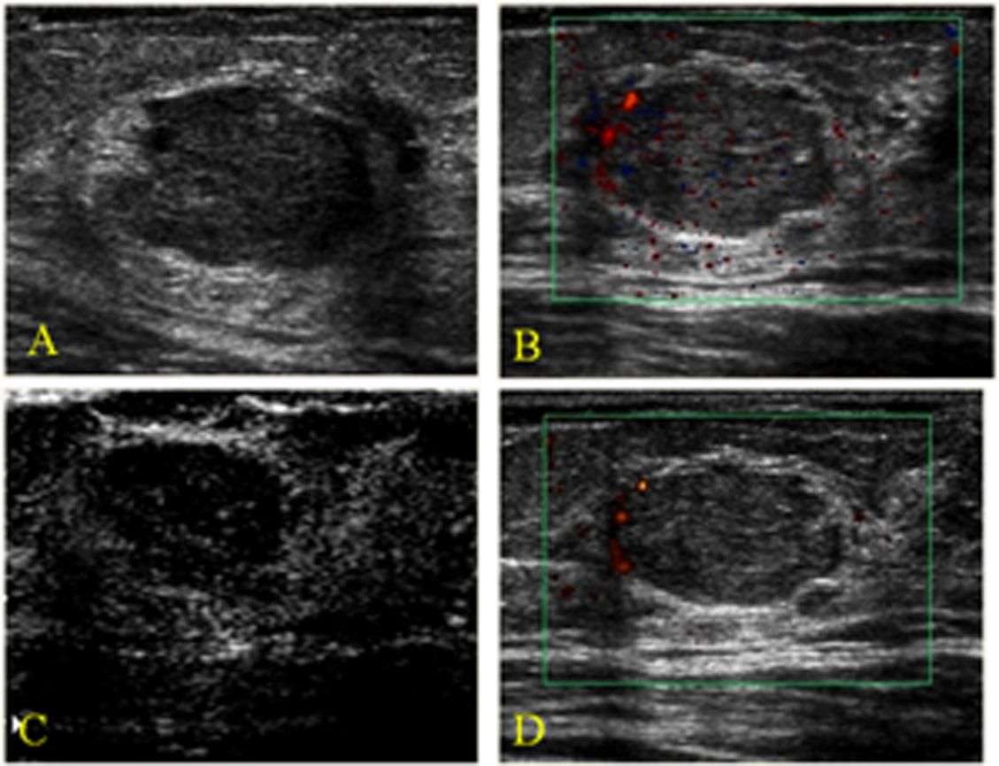
Ultrasound is useful because it can often tell the difference between fluid-filled masses like cysts and solid masses .
Ultrasound can also be used to help guide a biopsy needle into an area of the breast so that cells can be taken out and tested for cancer. This can also be done in swollen lymph nodes under the arm.
Ultrasound is widely available and is fairly easy to have done, and it does not expose a person to radiation. It also tends to cost less than other testing options.
You May Like: Can Lung Cancer Spread To The Breast
Benign Indeterminate And Malignant Nodules
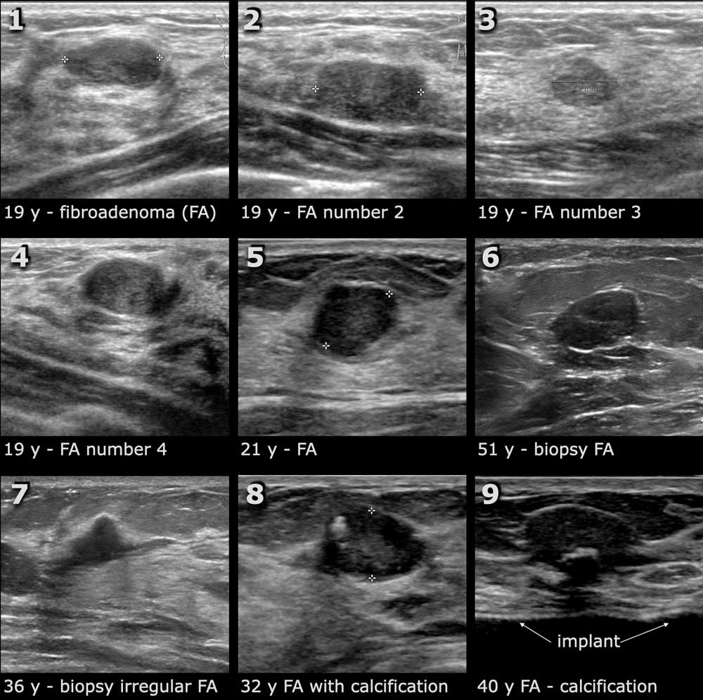
In a landmark study, Stavros et al established US criteria for characterizing solid breast masses. This work was facilitated by evolving technical improvements in US equipment that provided better resolution and images. They demonstrated that US may be used to accurately classify some solid lesions as benign, allowing follow-up with imaging rather than biopsy. They used high-resolution transducers, which were state-of-the-art at that time, and performed examinations in both radial and antiradial planes. The investigators also focused on the evaluation of suspected areas in the transverse and longitudinal planes.
Stavros et al proposed a US scheme for prospectively classifying breast nodules into 1 of 3 categories :
-
Indeterminate
To be classified as benign, a nodule had to have no malignant characteristics. In addition, 1 of the following 3 combinations of benign characteristics had to be demonstrated:
-
Intense uniform hyperechogenicity
-
Ellipsoid or wider-than-tall orientation, along with a thin, echogenic capsule
-
2 or 3 gentle lobulations and a thin, echogenic capsule
A nodule was classified as indeterminate by default if it had no malignant characteristics and none of the 3 benign characteristic combinations listed above.
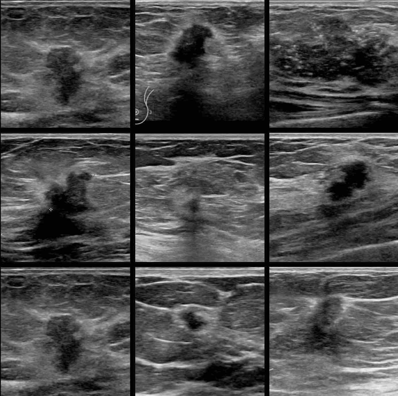
To be classified as malignant, a mass needed to have any of the following characteristics:
-
Spiculated contour
-
Branch pattern
-
Microlobulation
Whole Breast Ultrasound And Dense Breast Tissue
Studies have shown mammography combined with whole breast ultrasound may find slightly more breast cancers than mammography alone in women with dense breasts .
However, mammography plus whole breast ultrasound leads to more false positive results than mammography alone . False positive results must be checked to be sure theres no breast cancer. Follow-up tests, and sometimes a biopsy, are needed to check a false positive result.
There are no special screening guidelines for women with dense breasts . Whole breast ultrasound is not part of the NCCN or the ACS breast cancer screening recommendations for women at average risk or for women at higher than average risk .
Learn more about breast density and breast cancer risk.
Also Check: What Is Triple Negative Metastatic Breast Cancer
We Are Here For You All Herscan Breast Ultrasound Clinics Are Operating Private 20 Minute Appointments Provided For Each Customer
Our small, private clinics are kept at the highest hygienic standards possible, as we are actively following guidelines from the Centers for Disease Control and the World Health Organization. In addition, we are spacing out each appointment to limit the number of patients that are in each facility.
As we continue to monitor the COVID-19 situation very closely, we wanted to take a moment to address the steps we are taking to ensure the safety of our customers, staff and community. Please note, HerScan clinics take place in small, private environments that are less vulnerable than large clinics and hospitals.
All HerScan events will continue as scheduled. We remain vigilant, heeding all federal, state and local health advisories. Cleanliness standards are always a priority for the team at HerScan. However, due to concerns associated with COVID-19, we are taking additional steps so you are being served in the safest and most hygienic way possible.
To date, we have taken the following precautionary measures:
There is nothing more important to us than the health and safety of our customers and team members. We will continue to keep you informed and monitor this rapidly evolving situation.
Should I Have An Ultrasound Instead Of A Mammogram
In general, no. Its possible that breast ultrasounds may miss some smaller tumors that can be detected with mammography. In addition, ultrasounds are less accurate if you are overweight or have large breasts.
If you are pregnant, you should not have a mammogram. However, it is recommended that you discuss any breast changes with your doctor. Certain symptoms could indicate the need for an ultrasound.
Recommended Reading: How Common Is Breast Cancer In Teens
Tests To Determine Specific Types Of Treatment
You’ll also need tests that show whether the cancer will respond to specific types of treatment.
The results of these tests can give your doctors a more complete picture of the type of cancer you have and how to treat it.
In some cases, breast cancer cells can be stimulated to grow by hormones that occur naturally in your body, such as oestrogen and progesterone.
If this is the case, the cancer may be treated by stopping the effects of the hormones or by lowering the level of these hormones in your body. This is known as hormone therapy.
During a hormone receptor test, a sample of cancer cells will be taken from your breast and tested to see if they respond to either oestrogen or progesterone.
If the hormone is able to attach to the cancer cells using a hormone receptor, they’re known as hormone-receptor positive.
While hormones can encourage the growth of some types of breast cancer, other types are stimulated by a protein called human epidermal growth factor receptor 2 .
These types of cancers can be diagnosed using a HER2 test and are treated with medicine that blocks the effects of HER2. This is known as targeted therapy.
Want to know more?
Breast Ultrasound Vs Diagnostic Mammogram
Is a breast ultrasound better than a mammogram? Though a doctor might order both as a follow-up after an abnormal screening mammogram, there are several notable differences in the procedures. Some distinctions between the two are as follows.
- The imaging modality: Modality is the type of imaging used during a diagnostic test. Ultrasound uses sound waves to create images, while x-rays use radiation from electromagnetic waves to capture an image.
- The quality of images produced: Ultrasound and x-rays produce different types of pictures. Generally speaking, ultrasound images cant capture microcalcifications. Those tiny calcium deposits can often be some of the earliest signs of breast cancer. However, a mammogram can detect them.
- The reasons for the imaging: A physician might order either an ultrasound or a diagnostic mammogram to follow up on an abnormal screening mammogram. A breast ultrasound has uses beyond being a follow-up tool. In some cases, a doctor will perform a breast biopsy, collecting a sample of tissue from the breast and testing it for cancer. They might use ultrasound imaging to guide the needle to the correct area of the breast.
Read Also: What Does An Mri Show For Breast Cancer
Automated Whole Breast Ultrasound
Whole breast ultrasound exams are most often performed with a hand-held ultrasound device . The quality of the image can vary greatly, depending on the skill and experience of the person doing the exam.
Automated whole breast ultrasound is under study to see if it may help improve image quality . However, its not widely available or routinely used.
Calcifications On Breast Ultrasound Are Also Suspicious For Malignancy

Mammography is more sensitive than ultrasound when it comes to the detection of microcalcifications. Calcifications on a solid mass which appear punctate are highly suspicious of malignancy and will usually appear on ultrasound as bright, punctate foci.
Since malignant breast lesions are typically either intensely or mildly homogeneous hypoechoic solid masses, on ultrasound this provides a background which makes it easier to view calcifications sonographically. So, while calcifications are usually not seen on ultrasound, when they do appear vividly, it is highly suspicious for malignancy.
Don’t Miss: Does Breast Cancer Metastasis To Lung