Breast Cancer : Much Progress But Work Remains
Mention that statistic, and many women in the U.S. immediately know it refers to their lifetime risk of getting breast cancer.
Although the statistic may stir up anxiety, those diagnosed with breast cancer today have a more positive prognosis than ever, experts say. That’s due to better understanding of the disease, wider choices of treatments, and more individualized treatment designed to reduce the risk of recurrence and lessen side effects.
While breast cancer incidence has risen by 0.5% per year in recent years, and it remains the second leading cause of cancer death in women, outpaced only by lung cancer, there are now more than 3.8 million breast cancer survivors in the U.S.
If the disease is caught early, women with breast cancer have a survival rate of an astounding 99%, though that may dip to 28% if the cancer has spread.
But despite the progress, much work remains. Read on to see how far weve come in the fight against breast cancer — and what experts say needs to happen next.
Breast Cancer: Not a Single Disease
“Breast cancer is increasingly viewed as multiple different diseases,” says Harold J. Burstein, MD, a breast oncologist at the Dana-Farber Cancer Institute in Boston.
That discovery, in turn, has helped to individualize treatment and predict exactly how much treatment is needed for a specific patient, he and other experts say.
Molecular Diagnostics and ER-Positive Cancers
New Hope for HER2-Positive Cancers
Expanded Genetic Testing
Clean Margins In Breast Cancer Surgery
Hester Hill Schnipper, LICSW, OSW-CProgram Manager Emeritus, Oncology Social Work
MAY 13, 2019
Breast surgery is always a part of treatment for newly diagnosed non-metastatic breast cancer. Most often, the surgery, either a mastectomy or a lumpectomy/wide excision, happens as the initial treatment. It is then followed by radiation or chemotherapy, when appropriate, and hormonal therapy for estrogen receptor positive tumors. Sometimes women receive chemotherapy or hormonal therapy before surgery in this case, the medical treatment is called neo-adjuvant therapy.
Sometimes women have a mastectomy as the initial surgery . If a woman, instead, has a lumpectomy, the surgeon’s intention is to achieve clean margins. This means removing the malignant tumor with a surrounding area of healthy tissue. You likely have heard of circumstances when a woman needs to go back for a second or even third attempt to achieve these important clean margins. If several tries dont do it, the surgeon usually recommends a mastectomy.
Why are clean margins so important? Common sense tells us that removing the cancer is wise, and it seems sensible to take a little extra tissue to be sure all of the malignant cells are gone. A recent study supports this intent and gives us reasons and evidence why it is important.
Hearing that your surgeon got clean margins is a cause for relief and gratitude, and means that you can move forward to the rest of breast cancer treatment with optimism.
Experts Debate How To Achieve Both Clear Margins And The Best Cosmesis
IntroductionAnees B. Chagpar, MD, MSc, MA, MPH, FACS, FRCSBreast CenterSmilow Cancer Hospital at Yale-New Haven and Yale University School of MedicineIn a past column on ASCOconnection.org, I talked about a debate that had occurred in our tumor board in which a patient had a margin < 1 mm from ink. While technically negative, it was a little too close for comfort for me the surgeon whose case it was, however, argued based on evidence from the NSABP B-06 trial that if a tumor did not touch ink, outcomes were equivalent to the alternative of mastectomyat least for survival. It brought up how we interpret dataand the difference between what we know and what we think we know or as the comedian Stephen Colbert would put it, between truth and truthiness. We like to think that what we do is evidence-based, but we can almost always find data to support any position we wish to take.My two good friends, Dr. Mel Silverstein and Dr. Mike Dixon, have duked out the margins debate in many public forums and settle the score here once and for all. Here is what we know for sure: obtaining negative margins reduces local recurrence rates there is no consensus on what constitutes an adequate negative margin radiation therapy continues to play a role in breast-conserving surgery there are ways to take out large segments of breast tissue without compromising cosmesis and finally, for the record, Mel is not a Republican .
References
References
Recommended Reading: What Should You Eat If You Have Breast Cancer
Palpation Of Cancerous Masses
Cancerous masses in the breast are often very firm, like a rock or a carrot, and have an irregular shape and size. They are often fixedthey feel like they are attached to the skin or nearby tissue so that you can’t move them around by pushing on thembut can be mobile. They’re also not likely to be painful, though they can be in some cases.
On exam, other changes may be present as well, such as dimpling of the skin or an orange-peel appearance, nipple retraction, or enlarged lymph nodes in the armpit.
One type of breast cancer, inflammatory breast cancer, does not usually cause a lump but instead involves redness, swelling, and sometimes a rash on the skin of the breast.
New Evidence About Why Clear Margins In Breast Cancer Surgery Are Such Good News

- Date:
- Medical College of Georgia at Augusta University
- Summary:
- When a breast cancer tumor is removed with no signs of cancer left behind, it’s great news for patients, and now scientists have more evidence of why.
When a breast cancer tumor is removed with no signs of cancer left behind, it’s great news for patients, and now scientists have more evidence of why.
They’ve shown that once the breast tumor is successfully removed, the immune response turns its attention to destroying the tumor cells it has inevitably sent to nearby lymph nodes and organs like the lungs, says Dr. Hasan Korkaya, tumor biologist at the Georgia Cancer Center and Department of Biochemistry and Molecular Biology at the Medical College of Georgia at Augusta University.
The fact that metastasis did not occur in their model even months later, indicates the immune system destroyed the disseminated tumor cells rather than pushing them into a dormant state, Korkaya and his colleagues report in the journal Nature Communications.
In contrast, when the primary tumor was not completely removed, the immune system appears to begin to support the tumor, which grows back faster and bigger than the original mass, and its disseminated tumor cells survive.
“We wanted to see if we could mimic what was happening in the clinic,” says Korkaya, the study’s corresponding author.
Story Source:
Recommended Reading: Is Breast Cancer Caused By Smoking
New Guidelines Say Lumpectomy Margins Can Be Small As Long As Tumor Has No Ink On It
- Tags:Preparing for/Undergoing Surgery, Early-stage: Stage 0 — DCIS , Early-stage: Stage IA, Early-stage: Stage IB, Early-stage: Stage IIA, Early-stage: Stage IIB, Early-stage: Stage IIIA, Lumpectomy, Radiation to the Breast, Radiation After Surgery , Whole-Breast External Radiation, Ductal Carcinoma In Situ, Invasive or Infiltrating Ductal Carcinoma, and Invasive or Infiltrating Lobular Carcinoma
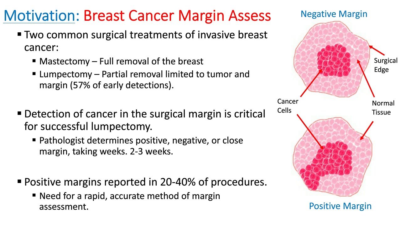
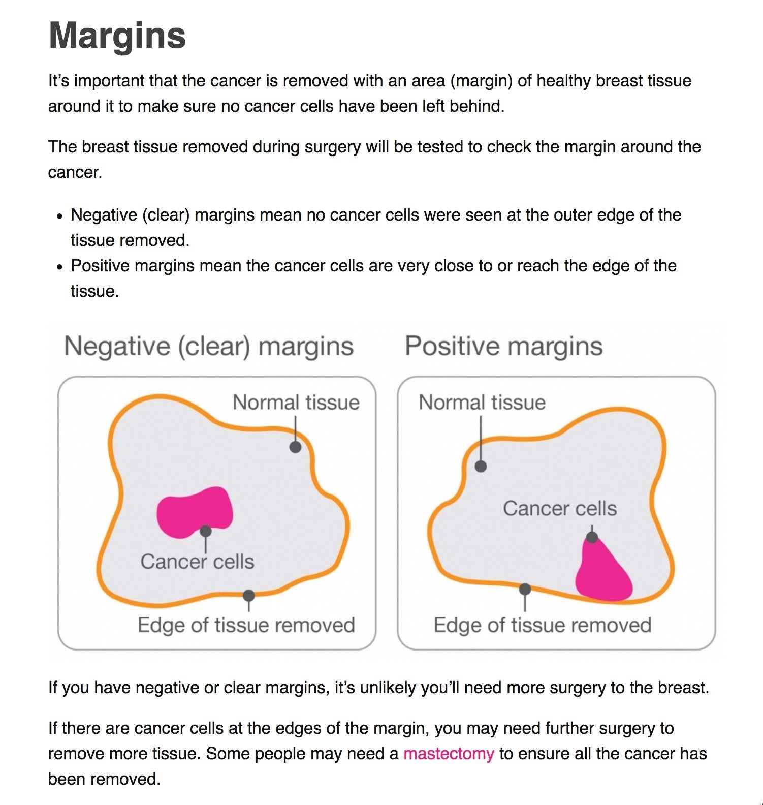
During lumpectomy, your surgeons goal is to take out all the breast cancer, plus a rim of normal tissue around it. This is to be sure all the cancer has been removed.
The tumor and surrounding tissue is rolled in a special ink so that the outer edges, or margin, are clearly visible under a microscope.
During or after surgery, a pathologist looks at the tissue thats been removed to make sure there are no cancer cells in the margin. A clear, negative, or clean margin means there are no cancer cells at the outer edge of tissue that was removed. A positive margin means that cancer cells come right out to the edge of the removed tissue and have ink on them. In some cases, a pathologist may classify the margins as close, which means that cancer cells are close to the edge of the healthy tissue, but not right at the edge and dont have ink on them.
The guidelines include several recommendations about margins after lumpectomy to remove early-stage breast cancer, including:
For more information on lumpectomy, including margins, visit the Breastcancer.org Lumpectomy pages.
Mastectomy And Tumor Margins
With a mastectomy, the whole breast is removed during surgery. Whether the margins contain cancer cells doesnt usually affect your treatment.
In rare cases after a mastectomy, the deep margin contains cancer cells. In these cases, more surgery and/or radiation therapy may be recommended.
With a nipple-sparing mastectomy, whether or not the nipple margin contains cancer cells can affect treatment. If the nipple margin contains cancer cells, more surgery and/or radiation therapy may be recommended.
Don’t Miss: What Is The Fish Test For Breast Cancer
Palpation Of Benign Breast Masses
In contrast to breast cancer tumors, benign lumps are often squishy or feel like a soft rubber ball with well-defined margins. They’re often easy to move around and may be tender.
Breast infections can cause redness and swelling. Sometimes it can be difficult to tell the difference between mastitis and inflammatory breast cancer, but mastitis often causes symptoms of fever, chills, and body aches, and those symptoms aren’t associated with cancer.
Optimizing Breast Cancer Therapy
As advances in breast cancer surgery and other modalities occur, we will continue to reevaluate whether we can de-escalate treatment approaches to lessen the burden of treatment for patients.
At MSK, we adopted the no ink on tumor consensus guideline early and conducted a study to confirm the benefits for our patients. We also pioneered the de-escalation of axillary dissection in women with invasive breast cancer and sentinel node metastasis following evidence that found no difference in overall survival or nodal recurrence between sentinel lymph node biopsy and complete axillary lymph node dissection.
The diagnosis and treatment of invasive breast cancer requires a collaborative, multidisciplinary approach. At MSK, the breast cancer team evaluates more than 4,500 new breast cancer cases and sees 3,300 surgical inpatients and outpatients annually. Our objective is to create the most effective individualized treatment plan for each patient to optimize outcomes, reduce the burden of treatment, and improve quality of life.
Monica Morrow, MD, FACS, Chief, Breast Service, Department of Surgery, and Anne Burnett Windfohr, Chair, Clinical Oncology, discuss MSKs evidence-based, leading-edge breast cancer surgical program.
Disclosure: Dr. Morrow has received honoraria from Genomic Health and Roche.
Don’t Miss: What Not To Eat With Breast Cancer
The Tissue Surrounding A Tumor And What It Means For Your Treatment
Jennifer Schwartz, MD, is a board-certified surgeon and Assistant Professor of Surgery at the Yale School of Medicine.
If you require a lumpectomy for breast cancer, your surgeon will remove the tumor and a border of tissue surrounding it called the surgical margin. A pathologist will then examine the tissue to determine if all the cancer cells in that area are gone or if further treatment is needed. If cancer cells are found anywhere between the tumor itself and the outer edge of the margin, additional surgery may be recommended.
Invasive Breast Cancer Margins: How Much Is Enough
As mentioned, the no ink on tumor guideline has led to a significant reduction in the use of additional surgery after an initial lumpectomy. At MSK, re-excision rates among women with invasive breast cancer declined significantly, from 21.4 percent before to 15.1 percent after early adoption of the guideline in January 2014. The use of BCT rose 13 percent over the same period.
In another study at MSK, we investigated the effect of margin width on LR in patients with triple-negative breast cancer who received BCT and analyzed the results for 535 cancers treated. Seventy-one cancers had margins less than or equal to 2 mm, and 464 had margins greater than 2 mm. Notably, there was no difference in the five-year LR rates between the smaller margin group and the larger margin group .
Read Also: What Is Treatment For Stage 2 Breast Cancer
Dcis Margins: How Much Is Enough
The ten-year cause-specific mortality rate for DCIS is under 1 percent after breast-conserving therapy, but optimizing local control is essential because half of all LRs are invasive cancers. The risk of LR for this patient population is affected by age, extent of disease, symptoms, presence of necrosis, margin width, and the use of adjuvant therapy.
Surveys of surgeons and radiation oncologists report a wide range of what constitutes an acceptable margin width, from no ink on tumor to greater than 1 centimeter. There is no uniform negative margin width reported in the literature that is associated with low LR risk in patients with DCIS treated with excision alone. Multiple factors inform the decision for re-excision or radiotherapy for DCIS, including the growth pattern of the lesion, margin width, the patients age, tumor size and grade, and the patients comfort with recurrence risk.
For women treated with excision alone, the goal is to remove all microscopic disease to minimize the risk of LR. For women undergoing lumpectomy and radiotherapy, an optimal margin leaves a subclinical volume of residual microscopic disease that can be controlled by radiotherapy.
No Association Of Positive Superficial And/or Deep Margins With Local Recurrence In Invasive Breast Cancer Treated With Breast

1Department of Surgery, Hanyang University College of Medicine, Seoul, Korea
2Division of Breast and Endocrine Surgery, Department of Surgery, Asan Medical Center, University of Ulsan College of Medicine, Seoul, Korea
3Department of Pathology, Asan Medical Center, University of Ulsan College of Medicine, Seoul, Korea
4Department of Oncology, Asan Medical Center, University of Ulsan College of Medicine, Seoul, Korea
5Department of Radiation Oncology, Asan Medical Center, University of Ulsan College of Medicine, Seoul, Korea
* Presented in part at the Global Breast Cancer Conference, Jeju, Korea, April 28-30, 2016.
You May Like: Does Having Breast Cancer Hurt
Dcis And Invasive Cancer: Which Guideline To Use
The invasive cancer margin guideline endorses no ink on tumor, while the DCIS guideline states that 2mm is an optimal margin. This raises the question of which guideline to apply in microinvasive carcinoma, or when DCIS occurs in association with invasive carcinoma and the DCIS component is in proximity to the margin. The margins consensus panel opted to draw the line for the DCIS guideline at microinvasive cancer, including this with DCIS because most of the lesion is comprised of DCIS, and because small retrospective studies suggest that the behavior of microinvasive carcinoma is more similar to DCIS than invasive cancer, and that the use of systemic therapy is more similar to that seen in DCIS. In contrast, invasive cancer with associated DCIS, whether an EIC or lesser amounts, should be managed according to the invasive guideline. In these cases, the biology of the invasive cancer is the primary determinant of outcome and the majority of patients will receive systemic therapy. Additionally, an EIC excised to clear margins does not increase LR,, although, as discussed previously, it is a potential marker for a heavier residual disease burden.
Breast Cancer Tumor Cells
Under the microscope, breast cancer cells may appear similar to normal breast cells or very little like breast cancer cells , depending on the tumor grade. Cancer cells differ from normal cells in many ways.
The cells may be arranged in clusters, and they may be seen invading blood vessels or lymphatic vessels. The nucleus of cancer cells can be striking, with nuclei that are larger, irregular in shape, and stain darker with special dyes. There are also often extra nuclei, rather than just one.
Don’t Miss: Does Alcohol Increase The Risk Of Breast Cancer
Suspicious Lesions And Lesions With A High Probability Of Malignancy
- Those that are benign and do not require any intervention
- Those that are probably benign and have a very low probability of being malignant, for which follow-up at short intervals is reasonable
- Those that are significant when they support or are supported by other indications of cancer
- Those that indicate moderate likelihood, should be considered suspicious, and should be biopsied
- Those that indicate a high probability of malignancy and require a biopsy
| Figure 16-1The classic description of breast cancer is an irregular mass with a spiculated margin. This irregular mass with spiculations was a small invasive ductal carcinoma. |
| Figure 16-2The morphology and margins of lesions are better seen on digital breast tomosynthesis. The first image is an enlarged view of a cancer that is hidden on the conventional mammogram by overlapping normal tissue. The lesion is more clearly seen in the second image that is a slice from the tomosynthesis study. |
| Figure 16-3 Although most breast cancers are fairly dense relative to fibroglandular breast tissue, this lesion is fairly low in x-ray attenuation, as seen on this craniocaudal projection and enlarged view . |
| Figure 16-4 Although most breast cancers are irregular in shape, some can be round and smoothly marginated, as was this invasive ductal carcinoma seen on this mediolateral oblique and craniocaudal mammogram. |
| Figure 16-11 Cancer may be hidden in dense fibroglandular tissue, as in this craniocaudal mammogram. |