Implications For The Treatment Of Estrogen
Our results also demonstrate that PGE2 suppresses p53 expression in ASCs. Whether this also occurs in breast cancer cells remains to be determined however, previous studies have demonstrated that overexpression of COX2, the rate-limiting step in prostaglandin synthesis, is associated with the repression of p53 target genes in normal human mammary epithelial cells . Inhibition of COX2 using nonsteroidal anti-inflammatory drugs is associated with a decreased breast cancer risk and has been proposed as a means of breast-specific aromatase inhibition. Therefore, COX2 inhibitors may be useful for the treatment of estrogen-dependent breast cancers by restoring p53 expression and inhibiting estrogen production and cancer cell growth.
P53 Accumulation In Breast Cancer Cells With Missense Mutation Of Tp53 Gene
We first analyzed p53 expression by immunohistochemistry in a selection of cancer cell lines using formalin-fixed, paraffin-embedded cell block materials . Negative or week nuclear staining in only a few cells was observed in H1299 p53 null cells and in H2228, Lovo, Hela and MCF-7 cells without the TP53 gene mutation . In contrast, strong nuclear staining in all cells was seen in T47D, SK-BR-3, BT-474, MDA-MB-231 and MDA-MB-468 cells with missense mutations of the TP53 gene .
Figure 2
| 82.8 ± 23.9 | 80.6 ± 32.8 |
- *P < 0.05 is considered significant. ER, estrogen receptor HER2, human epidermal growth factor receptor type 2 LI, labeling index PR, progesterone receptor.
Oncogenes Mdm2 And P53
In the frequency of its disruption in human cancer, the CDKN2A gene, located at 9p21, is second only to Tp53 . In fact, this locus encodes two proteins translated in alternate reading frames: P16INK4A, a tumour suppressor, is a cyclin-dependent kinase inhibitor that acts upstream of retinoblastoma protein to promote cell-cycle arrest P19ARF is more related to p53 activity. P16INK4A and
P19ARF are often co-deleted in tumour cells, as notably observed in the widely used, wild-type p53 MCF-7 breast cancer cell line , but mice lacking P19ARF alone are highly susceptible to breast cancer , thus underlining its importance.
P19ARF activates p53 by sequestering MDM2 into the nucleolus, thus preventing it degrading p53. The P19ARF-p53 axis is critical for eliminating potential tumour cells containing deregulated oncogene expression. The adenoviral proteins E1A and MYC, when over-expressed, may promote apoptosis through p53 activation. By the same pathway, V-Ha-ras Harvey rat sarcoma viral oncogene homologue may induce cell senescence. It has been shown that P19ARF is strictly required to mediate these effects on p53 . P19ARF may also mediate the positive effects of beta-catenin on p53 activity .
Also Check: Anne Hathaway Breast Cancer
Can I Have Tp53 Genetic Testing
In the past, the diagnosis of LFS was made by clinical criteria, meaning it was based on the signs and symptoms the patient and family had. Now, genetic testing is available for people to learn whether they carry a copy of the TP53 mutation before any physical signs of LFS appear. The decision to test is highly personal. People considering TP53 genetic testing are strongly encouraged to receive professional genetic counseling first, so they can gain the knowledge they need to make an informed decision. This can also help with the serious emotional effects that may occur when people learn that they are a carrier. Genetic counseling, as a part of considering genetic testing, is important not only for the patient but also for that persons relatives.
Testing a child in a family with LFS is a complex situation since the decision to do testing must made by the childs parents, with the help of medical experts. However, since cancers occur often among children in families with LFS, testing at-risk children, rather than delaying testing until young adulthood, must also be strongly considered when the goal is to find LFS-related cancers early and treat them more effectively.
If genetic testing shows that a person has a TP53 mutation, this may mean that their doctor could recommend surveillance, which means being monitored regularly for LFS-related types of cancer. This is an in-depth, lifelong process. More about the surveillance process is outlined below.
Experimental Analysis Of P53 Mutations

Most p53 mutations are detected by DNA sequencing. However, it is known that single missense mutations can have a large spectrum from rather mild to very severe functional affects.
The large spectrum of cancer phenotypes due to mutations in the TP53 gene is also supported by the fact that different isoforms of p53 proteins have different cellular mechanisms for prevention against cancer. Mutations in TP53 can give rise to different isoforms, preventing their overall functionality in different cellular mechanisms and thereby extending the cancer phenotype from mild to severe. Recents studies show that p53 isoforms are differentially expressed in different human tissues, and the loss-of-function or gain-of-function mutations within the isoforms can cause tissue-specific cancer or provides cancer stem cellpotential in different tissues. TP53 mutation also hits energy metabolism and increases glycolysis in breast cancer cells.
It was initially presumed to be an oncogene due to the use of mutated cDNA following purification of tumor cell mRNA. Its role as a tumor suppressor gene was revealed in 1989 by Bert Vogelstein at the Johns Hopkins School of Medicine and Arnold Levine at Princeton University.
In 1993, p53 was voted molecule of the year by Science magazine.
Don’t Miss: Did Anne Hathaway Have Breast Cancer
P53 As A Metabolic Checkpoint
Emerging evidence suggests that p53 is also involved in the regulation of metabolism and cell homeostasis without causing cell-cycle arrest or apoptosis . For example, nutrient deficiency leads to the activation of p53 through direct phosphorylation at Ser15 by AMP-activated protein kinase , a key regulator of cell metabolism . p53 also regulates metabolism and cell homeostasis in normal cells and tissues. Lipin1 is a recently identified p53 target gene that regulates the expression of genes involved in fatty acid oxidation through PPAR . Under nutrient/glucose deprivation conditions, p53 is upregulated by AMPK and stimulates Lipin1 and malonyl-CoA decarboxylase expression, leading to an increase in fatty acid oxidation . This allows cells to use fatty acids as an alternative energy source. Reciprocally, p53 also promotes the expression of AMPK, leading to the negative regulation of mTOR . The PI3K/Akt/mTOR pathway can suppress apoptosis and stimulate proinflammatory gene expression, which in turn promotes cancer growth and progression . Negative regulation of mTOR is also observed in autophagy, which can be induced by starvation and metabolic stresses.
In summary, a number of genes are regulated by p53 to maintain metabolism and energy homeostasis in cells/tissues under normal and stressed physiologic conditions. Changes in tumor cell metabolism are now recognized as a hallmark of cancer and p53 is integral to this process.
Induction Of Genetic Instability
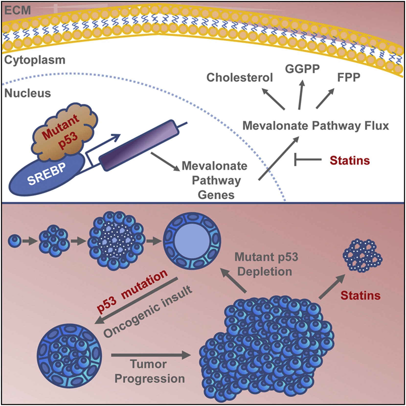
As the Guardian of the genome, the fundamental goal of WT p53 is to maintain genetic stability by preventing the passage of genetic mutations to daughter cells . While p53 null cells still retain certain levels of checkpoint and DNA repair capacities, cells harboring p53 mutant proteins showed a dramatic higher level of genomic instability such as interchromosomal translocations and aneuploidy, indicating the oncogenic GOF activity of p53 mutants . These variations largely contribute to genetic diversity that expedites malignant tumor development. Mechanistically, the common p53 mutants can disrupt the earliest stage of DNA double-stranded break damage responses by interacting with the nuclease Mre11 to suppress the recruitment of Mre11/Rad50/NBS1 complex to the site of DNA DSB damage, leading to inactivation of ATM, the key DNA DSB damage sensor, and the resultant G2/M checkpoint impairment . Mutp53 can also induce genomic abnormality by inactivating DNA replication process. For example, some mutp53 proteins activate cyclin A to promote the formation of DNA replication origin and the intra-S phase checkpoint kinase CHK1 to stabilize the replication forks, facilitating the duplication of aberrant genomic DNAs .
Recommended Reading: Is Breast Cancer Curable In The 3 Stage
P53 Cooperates With Many Pathways Involved In Mammary Tumorigenesis
In addition to breast cancer susceptibility genes, p53 has been studied for cooperativity with pathways activated in sporadic breast cancer. Transgenic mice overexpressing oncogenes involved in human breast cancer, such as HER-2/Neu, Wnt1 and c-myc, have been generated that develop a high incidence of sporadic mammary carcinomas. By crossing these mice with Trp53 mutant mice or null mice to generate double-transgenic mice, it has been possible to directly demonstrate pathway cooperativity . For example, HER-2, a member of the epidermal growth factor receptor family, is overexpressed in approximately 20% of human breast cancers and is associated with a poor prognosis .
Transgenic mice expressing wild-type rat neu under the MMTV promoter develop mammary tumors with an average latency of 234 days that have a high frequency of missense mutations in Trp53. The cooperativity between the Neu and p53 pathways was clearly demonstrated by introducing the mutant p53 transgene, p53-R172H, into these mice, which reduced tumor latency to 154 days . This study in transgenic mice has thus demonstrated strong cooperativity between two genetic lesions common in human breast cancer, and has demonstrated that p53 mutation is an important event in Neu-mediated oncogenesis.
Depletion Of Mtp53 R273h Modulates Parp Localization And Parp Enzymatic Activity
PARP family enzymes are critical for a number of biological processes, including regulating transcription, DNA replication, and DNA repair. Moreover, PARP catalyzes the transfer of ADP ribose to target proteins. Interestingly, we observed through SILAC experiments that the depletion of mtp53 in MDA-468.shp53 cells increased the level of PARP1 in the cytoplasmic fraction and decreased the level of PARP1 in the chromatin fraction . The LC-MS/MS high-quality data and the reduction of PARP in the nuclear fraction was exemplified by examining the 3+ ions for the relative H/L abundance of m/z for PARP1 . The MS data also demonstrated that depletion of mtp53 correlated with a reduction of PCNA in the cytoplasmic and chromatin fractions .
Recommended Reading: What Is Stage 3a Breast Cancer
Questions To Ask The Health Care Team
If you are concerned about your risk of cancer, talk with your health care team. It can be helpful to bring someone along to your appointments to take notes. Consider asking your health care team the following questions:
-
What is my risk of developing cancer?
-
Should I receive a risk assessment, genetic counseling, and discuss genetic testing? If so, how can I do that?
-
What can I do to reduce my risk of cancer?
-
What are my options for cancer screening and prevention?
If you are concerned about your family history and think your family may have LFS, consider asking the following questions:
-
Does my family history increase my risk of cancer?
-
Could my family have LFS?
-
Will you refer me to a hereditary cancer clinic to meet with a genetic counselor and other genetics specialists?
-
Should I consider genetic testing?
Tp53 Gene And Its Mutations In Spontaneous Breast Cancer
According to the current release of the International Agency for Research on Cancer TP53 database , included in COSMIC, ~70% of the breast cancer alterations in TP53 are missense mutations . This proportion, as well as the spectrum of mutated codons in the gene , reflect the p53 mutational pattern of other tumors . A noteworthy difference is codon 220, which is the fourth most frequent missense mutation in breast cancer , whereas it ranks seventh in other cancers . Another such overrepresentation is codon 163 . Although no explanation of these differences has been provided, geographic or ethnical characteristics have been suggested to influence the occurrence of specific mutations, possibly due to the link to environmental mutagens . Associations of TP53 mutation with breast-cancer-predisposing BRCA1/2 germline mutations have been also found, probably favored by a bias in the dysfunctional DNA repair mechanisms . In sporadic breast cancers, high TP53 mutation frequencies have been significantly associated with two polymorphisms: the homozygous Arginine at codon 72 of p53 and the presence of the highly active allelic variant G of glutathione-S-transferases . Importantly, differences have been found in the specific TP53 mutation occurrence in breast cancer types and grades, as well as in the survival of patients bearing particular hotspot mutations .
Recommended Reading: Can Getting Hit In Your Breast Cause Cancer
Changes In P53 Coactivators
In addition to proteins such as ATM, ATR and Chk2 that regulate the stability and function of p53 through phosphorylation, a second, functionally distinct, group of proteins is now emerging that appear to operate as cofactors stimulating one or more of the wild-type properties of p53. One such family with possible involvement in breast cancer is the apoptosis stimulating protein of p53 . Two members of this family , encoded by separate genes, have recently been described .
Expression of either ASPP1 or ASPP2 stimulates the proapoptotic function of wild-type p53 by increasing p53-dependent induction of apoptotic effectors such as Bax and PIG3, whereas expression of nonapoptotic proteins such as p21Waf1 was much less affected. In primary breast cancers lacking p53 mutation, expression of both ASPP1 and ASPP2 was reduced. These observations are supported by an earlier report that, using microarray methodology, also identified p53 BP2 downregulation in breast cancer . Taken together, these studies suggest that downregulation of ASPP proteins attenuates p53-dependent apoptosis, thus conferring a selective advantage to breast carcinomas with intact p53.
Changes In Upstream Regulators Of P53

Aside from the two well-recognised breast cancer susceptibility genes BRCA1 and BRCA2, it is an attractive hypothesis that mutations in other genes with lower penetrance may account for a significant proportion of hereditary breast cancers. One such gene may be ATM, the gene mutated in ataxia-telangectasia . The link between ATM and p53 was suggested in early studies revealing defective induction of p53 following irradiation of A-T cells. It has subsequently been established that phosphorylation of both p53 and BRCA1 in response to -irradiation occurs via ATM.
A-T patients have a high incidence of cancer, and some develop breast carcinomas . A recent study examined the entire ATM coding sequence in a large series of breast cancer patients and identified heterozygosity for truncating mutations in approximately one in 50 patients, consistent with the hypothesis that A-T heterozygotes are more common in breast cancer patients than in the general population . It has been hypothesised that inactivation of ATM may be an alternative to p53 mutation in leukaemia. There is evidence that low or absent expression of ATM occurs commonly in sporadic breast cancer . Interestingly, some cancers in this study had both low ATM expression and p53 mutation, suggesting that the inactivation of the two genes is not necessarily exclusive.
Don’t Miss: Can Asbestos Cause Breast Cancer
Mechanisms Of P53 Apoptosis Vs Growth Arrest P53 Apoptotic Co
Apoptosis appears as the critical function of p53 in tumour suppression . The choice between growth arrest and apoptosis likely involves the complex interplay of numerous factors.
According to a quantitative model, genes involved in growth arrest contain high-affinity p53 binding sites in their promoter, while low-affinity sites are present in the promoter of apoptosis-related genes . This is in line with observations that increased levels or activity of p53 can lead to the onset of apoptosis, presumably by achieving a certain threshold level. Moreover, p53 mutants with marginally altered conformations retain sufficient activity to induce growth arrest but not apoptosis, presumably because they can still interact only with high-affinity sites. However, despite the degenerative nature of p53 binding sequences, the apoptotic targets of p53 do not necessarily contain low-affinity promoters. For example, chromatin immunoprecipitation experiments have revealed that the apoptotic gene BBC3 contains high-affinity p53 binding sites . The quantitative model is thus not sufficient.
As mentioned above, phosphorylation of the p53 residue Ser46 plays an important role in permitting the apoptotic function of the protein. The interaction between p53DINP1 and Ser46 may allow this phosphorylation.
Germline Vs Somatic Mutations
Germline mutations are the type of mutations people may be concerned with when wondering if they have a genetic predisposition to cancer. The mutations are present from birth and affect every cell in the body. Genetic tests are now available that check for several germline mutations that increase cancer risk, such as mutated BRCA genes. Germline mutations in the TP53 gene are uncommon and associated with a specific cancer syndrome known as Li-Fraumeni syndrome.
People with Li-Fraumeni syndrome often develop cancer as children or young adults, and the germline mutation is associated with a high lifetime risk of cancers, such as breast cancer, bone cancer, muscle cancer, and more.
Somatic mutations are not present from birth but arise in the process of a cell becoming a cancer cell. They are only present in the type of cell associated with the cancer , and not other cells in the body. Somatic or acquired mutations are by far the most common type of mutation associated with cancer.
Also Check: How To Cure Breast Cancer With Baking Soda
Dna Damage And Repair
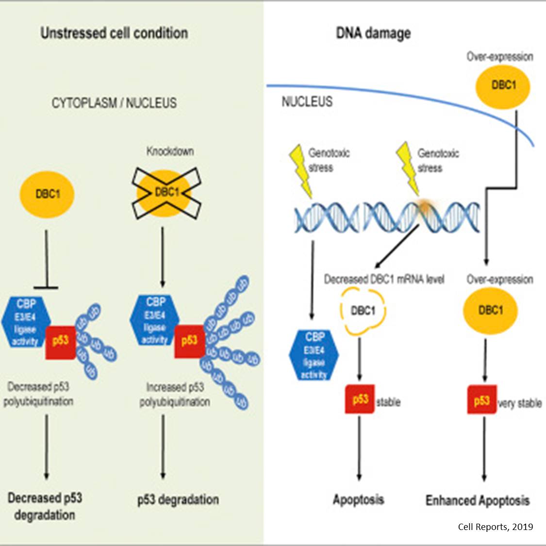
p53 plays a role in regulation or progression through the cell cycle, apoptosis, and genomic stability by means of several mechanisms:
- It can activate DNA repair proteins when DNA has sustained damage. Thus, it may be an important factor in aging.
- It can arrest growth by holding the cell cycle at the G1/S regulation point on DNA damage recognitionif it holds the cell here for long enough, the DNA repair proteins will have time to fix the damage and the cell will be allowed to continue the cell cycle.
- It can initiate apoptosis if DNA damage proves to be irreparable.
- It is essential for the senescence response to short telomeres.
p53 pathway
WAF1/CIP1 encoding for p21 and hundreds of other down-stream genes. p21 binds to the G1–S/CDK complexes inhibiting their activity.
When p21 is complexed with CDK2, the cell cannot continue to the next stage of cell division. A mutant p53 will no longer bind DNA in an effective way, and, as a consequence, the p21 protein will not be available to act as the “stop signal” for cell division. Studies of human embryonic stem cells commonly describe the nonfunctional p53-p21 axis of the G1/S checkpoint pathway with subsequent relevance for cell cycle regulation and the DNA damage response . Importantly, p21 mRNA is clearly present and upregulated after the DDR in hESCs, but p21 protein is not detectable. In this cell type, p53 activates numerous microRNAs that directly inhibit the p21 expression in hESCs.