Lowering The Risk Of Breast Cancer
Some hormonal therapy drugs can reduce the risk of breast cancer developing in women with LCIS. Your doctor or specialist nurse may talk to you about taking a hormonal therapy if it is an option for you. It is not clear how much the reduction in risk outweighs the side effects of hormonal therapy drugs.
You might have a pattern of breast cancer in your family. For example, there may be a number of family members who have been diagnosed with breast cancer. If this is the case, you may be referred to a genetics clinic to see a specialist. At the clinic, you have a risk assessment and genetic counselling.
Some women who are assessed as having a high risk of breast cancer, may think about having surgery to remove both breasts removed. This is to reduce the risk of breast cancer developing. It is called risk-reducing breast surgery.
Talk to your doctor or specialist nurse if you are worried about your family history or risk of developing breast cancer.
Lobular Carcinoma In Situ And Ductal Carcinoma In Situ Are Abnormalities That Doctors Call Stage Zero Breast Cancer Women With Either Of These Diagnoses Often Ask Us Do I Have Breast Cancer
Despite the fact that its name includes the term “carcinoma,” LCIS is not a true breast cancer. Rather, LCIS is an indication that a person is at higher-than-average risk for getting breast cancer at some point in the future. For this reason, some experts prefer the term lobular neoplasia instead of lobular carcinoma. A neoplasia is a collection of abnormal cells. LCIS is restricted to the lobules.
DCIS is the most common kind of carcinoma in situ. In DCIS, cancer cells are only in the ductal walls. If DCIS is not treated, it will likely grow into an invasive cancer. Here is a side-by-side comparison of the two conditions:
What Should You Tell The Patient And The Family About Prognosis
Ductal carcinoma in situ
DCIS is curable and the prognosis is excellent. Because it is non-invasive, the tumor does not have the capacity to spread distantly. In rare cases, particularly with large DCIS lesions greater than 4cm, metastatic disease to lymph nodes or distant sites may occur. This is likely due to unidentified areas of micro-invasion that are not seen during pathologic sampling.
The risk of recurrence of in situ or invasive cancer in the ipsilateral breast is related to the extent of surgical treatment:
-
less than 1% with total mastectomy
-
7-11% at 10 years with breast conserving surgery and radiation.
For patients with Van Nuys scores of 4-6, recurrence risk is 6% or less. The most serious adverse consequence of recurrence is that the lesion may be invasive 50% of the time when it recurs, which then exposes the patient to the risk of distant disease.
Lobular carcinoma in situ
LCIS is a risk factor for the development of subsequent breast cancer in either breast. The risk of subsequent invasive breast cancer ranges from 7% to 17% at 10-15 years after LCIS diagnosis. Slightly more than half of these cases occur in the contralateral breast. This risk can be reduced by endocrine systemic therapy, with tamoxifen for pre-menopausal women, and tamoxifen, raloxifene or exemestane for post-menopausal women.
Don’t Miss: Stage Iv Breast Cancer Symptoms
Breast Cancer Risk After Lcis Diagnosis
It is estimated that women with LCIS have 7 to 12 times the risk of developing breast cancer compared to women without LCIS. Most breast cancers involve the milk ducts and not the lobules, and this tendency does not change if you have had LCIS.
LCIS is a sign of an increased predisposition to breast cancer, but not necessarily lobular breast cancer. LCIS is not considered a precursor of breast cancer, and the cells do not change or become cancer cells.
Classification Of Lobular Carcinoma In Situ

Currently, histological features guide classification of LCIS lesions. The three main histological sub-classifications of LCIS are classical , florid and pleomorphic , and these entities can be found to coexist.
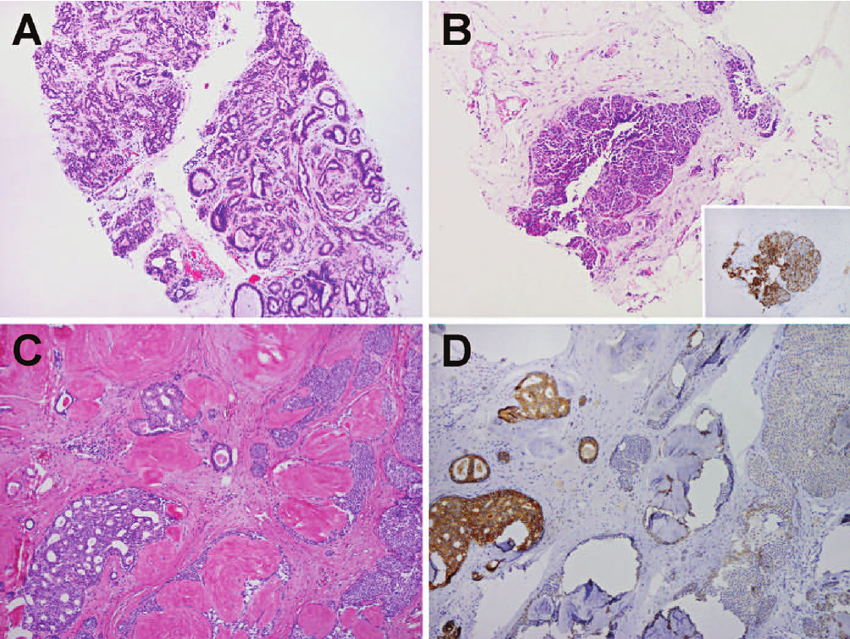
Histologically, CLCIS is characterized by a monomorphic population of small round cells with a ring of clear cytoplasm . Cells within the lesion are loosely adherent, filling the lumen of the acini and distending the TDLU, yet they maintain the architecture of the lobules with an intact basement membrane and myoepithelial cell layer . Mitotic figures and necrosis, as well as calcifications, are not common in CLCIS. Pagetoid spread, in which neoplastic cells extend along the mammary ducts, is frequently observed. There are two categories of CLCIS, type A and type B . Type A CLCIS is generally low grade, with small nuclei and inconspicuous nucleoli. Type B CLCIS is composed of cells with larger nuclei and small nucleoli. CLCIS tends to be positive for estrogen receptor and progesterone receptor , and negative for HER2.
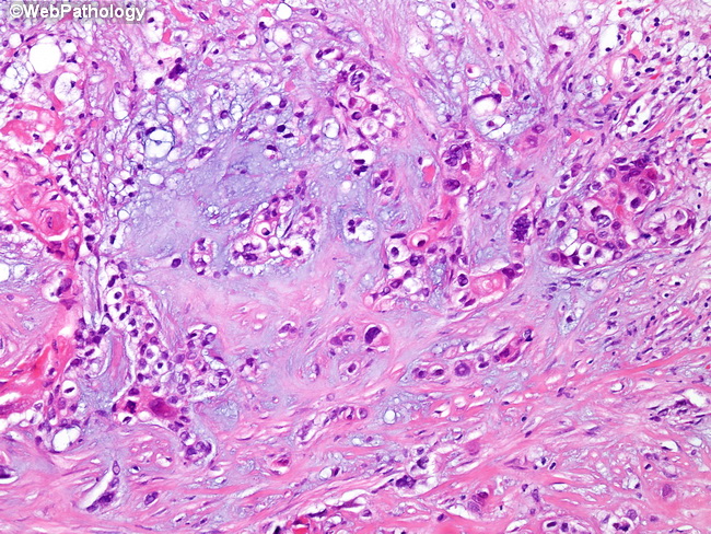
FLCIS is a comparatively more rare lesion, histologically characterized by massive expansion of the involved TDLUs, often associated with necrosis and calcifications. Morphologically, it resembles solid-type DCIS. The lesion is frequently associated with ILC, supporting FLCIS as a precursor of ILC . FLCIS shows more genetic instability than CLCIS, including a higher fraction of genomic alterations and breakpoints .
Also Check: Anne Hathaway’s Breasts
What Is Lobular Carcinoma In Situ
Lobular carcinoma in situ is a rare condition that happens when you have abnormal cells in your lobules the glands that produce breast milk. These abnormal cells are in situ, meaning they havent spread to surrounding breast tissue.
Lobular carcinoma in situ isnt breast cancer. But its a marker or indication that you have greater risk for developing breast cancer than someone who doesnt have LCIS. If you have lobular carcinoma in situ, you are 10 times more likely to develop breast cancer than someone who doesnt have LCIS.
What About Mastectomy To Prevent Future Breast Cancer
More than 20 years ago, when breast conditions like these were diagnosed, they were often treated with mastectomy, surgery which completely removes the affected breast. Sometimes a healthy second breast was also removed , even when there was no sign of cancer or other abnormalities in the other breast.
Today, thanks to advances in scientists understanding of breast cancer and of these other conditions, along with the development of better diagnostic, surgical, and treatment techniques, mastectomy is often unnecessary. In fact, we now know that a less radical treatment or no treatment is just as effective. The latest research indicates that women who undergo lumpectomy and radiation rather than mastectomy tend to live longer. Except in unusual circumstances, mastectomy does not increase survival time for these conditions, and the risks of mastectomy usually outweigh any benefits.
All articles are reviewed and approved by Dr. Diana Zuckerman and other senior staff.
Related Content:
Read Also: How Many Men Have Breast Cancer
How Is Plcis Treated
If the biopsy shows PLCIS, your doctor may suggest an operation to remove the area with a margin of healthy breast tissue. This is because of the higher breast cancer risk with this type of lobular neoplasia. The operation will show if there are any cancer cells in the tissue, and whether all the PLCIS has been removed.
Often, PLCIS is treated in the same way as ductal carcinoma in situ , which is a type of breast cancer. Radiotherapy or hormone therapy may be recommended.
What Does It Mean If My Diagnosis Says Lobular Carcinoma In Situ Lobular Neoplasia Or In
There are 2 main types of in-situ carcinoma of the breast: ductal carcinoma in situ and lobular carcinoma in situ . They are diagnosed based on how the cells and tissue look under the microscope, and are sometimes both found in the same biopsy.
LCIS and a condition called atypical lobular hyperplasia are both considered lobular neoplasia.
In-situ carcinoma with duct and lobular features means that the in-situ carcinoma looks like DCIS in some ways and LCIS in some ways , so the pathologist cant call it one or the other.
Lobular carcinoma in situ is a type of in-situ carcinoma of the breast. While DCIS is considered a pre-cancer, it is unclear whether LCIS is definitely a pre-cancer or if it is just a general risk factor for developing breast cancer. This is because LCIS rarely seems to turn into invasive cancer if it is left untreated. Women with LCIS do have a higher risk of getting breast cancer, but the cancer occurs just as often in the opposite breast . Because it isn’t clear if LCIS is a pre-cancer, many doctors prefer to use the term lobular neoplasia instead of lobular carcinoma in situ.
If LCIS is found in an excision biopsy, it does not need further treatment. Because it increases the risk of a later cancer, your doctor might discuss taking medicine to lower your risk of breast cancer.
Also Check: Stage 3 B Breast Cancer
What Should The Initial Definitive Therapy For The Cancer Be
Ductal carcinoma in situ
Surgical procedures
DCIS may be treated with breast conserving surgery or mastectomy with or without immediate reconstruction. The decision for type of surgery is based on the extent of the lesion relative to breast size, whether adequate clear margins can be achieved with partial mastectomy, ability to receive radiation therapy, and patient preference.
With mastectomy, skin sparing techniques can be utilized to facilitate reconstruction with excellent cosmesis. However, it is important that anterior margins of the mastectomy are clear of calcifications and tumor. for thin patients with large areas of microcalcifications, skin sparing may result in a positive margin on the mastectomy specimen if skin over these calcifications is not taken in the resection.
Radiation Therapy
For DCIS treated with breast conserving surgery, radiation is indicated in most circumstances to reduce risk of local recurrence. Clinical trials have demonstrated a 50-60% reduction in local recurrence. However, for certain tumors, that absolute benefit is low.
The following factors should be considered in determining the role of radiation therapy:
-
size of the DCIS
-
tumor grade and extent of comedonecrosis
-
width of margins
-
patient age
Table II.
Van Nuys Prognostic Index
Surgical margins
Radiographic evaluation
Lymph node evaluation
Circumstances in which sentinel node biopsy should be considered even with breast conserving surgery include:
Endocrine therapy
The Breast Cancer Prevention Trial
Because of the observation that women with breast cancer treated with Nolvadex® had a lower risk of developing a new breast cancer in their unaffected breast, many doctors felt that Nolvadex® may actually be able to prevent breast cancer from occurring. The results of the National Cancer Institute clinical study evaluating Nolvadex® attempted to address whether Nolvadex® could decrease the number of breast cancers and thereby decrease the number of women dying from breast cancer. Other goals of the study were to determine whether Nolvadex® could also decrease the number of heart attacks and bone fractures, as well as determine whether Nolvadex® has any detrimental side effects.
Also Check: Did Anne Hathaway Have Cancer In Real Life
Are You Sure Your Patient Has Stage 0 Breast Cancer What Should You Expect To Find
Ductal carcinoma in situ
DCIS is usually asymptomatic and identified first with an abnormal mammogram. In modern times, DCIS will rarely present as a palpable mass. A spontaneous nipple discharge which is bloody, pink tinged, clear or serous in nature may be a presenting symptom.
Lobular carcinoma in situ
LCIS is usually asymptomatic and will be found as an incidental finding on biopsy of the breast for other findings. LCIS is found in approximately 1% of all excisional breast biopsy specimens. About 80% of LCIS occurs in pre-menopausal women .
Lobular Carcinoma In Situ

Lobular carcinoma in situ is a type of breast change that is sometimes seen when a breast biopsy is done. In LCIS, cells that look like cancer cells are growing in the lining of the milk-producing glands of the breast , but they dont invade through the wall of the lobules.
LCIS is not considered to be cancer, and it typically does not spread beyond the lobule if it isnt treated. But having LCIS does increase your risk of developing an invasive breast cancer in either breast later on, so close follow-up is important.
LCIS and another type of breast change are types of lobular neoplasia. These are benign conditions, but they both increase your risk of breast cancer.
Read Also: Estrogen Dependent Breast Cancer
Treatment Choices For Dcis
DCIS patients have three surgery choices. They are 1) lumpectomy followed by radiation therapy 2) mastectomy or 3) mastectomy with breast reconstruction surgery. Most women with DCIS can choose lumpectomy.
Lumpectomy means that the surgeon removes only the cancer and some normal tissue around it. This kind of surgery keeps a womans breast intact looking a lot like it did before surgery. Under most circumstances, mastectomy does not increase survival time for women with DCIS, and would only be considered under unusual circumstances, such as cases where the breast is very small or the area of DCIS is very large. For women who undergo mastectomy, reconstruction can replace the breast lost to cancer. However, there is some evidence that women with DCIS who undergo mastectomy do not live as long as those who undergo lumpectomy.
Radiation therapy is often recommended for almost all women with DCIS after lumpectomy. This type of treatment is very important because it could keep more DCIS or invasive cancer from developing in the same breast. However, DCIS patients who choose lumpectomy live just as long whether they undergo radiation or not. DCIS patients who undergo a single mastectomy or double mastectomy do not live any longer than DCIS patients who undergo lumpectomy.
Active surveillance is gaining attention as an option for women with DCIS. Active surveillance consists of regular mammography screening to make sure the DCIS does not develop into breast cancer.
Lobular Carcinoma In Situ An Overview
Lobular carcinoma in situ is an area of abnormal cell growth that occurs in the lobules of the breast that increases an individual’s risk of developing invasive breast cancer. Lobules are the milk-producing glands at the end of breast ducts. Individuals diagnosed with LCIS tend to have more than one lobule affected.
LCIS is not a true breast cancer but an indication that an individual is at higher-than-average risk for developing an invasive breast cancer at some point in the future.
LCIS is multifocal, meaning that multiple lobules have areas of abnormal cell growth inside them in more than half of women diagnosed with the condition. In about one-third of women with LCIS, the other breast will be affected
LCIS is usually diagnosed before menopause, most often between the ages of 40 and 50. Less than 10% of women diagnosed with LCIS have already gone through menopause. LCIS is extremely uncommon in men.
Recommended Reading: Signs Of Breast Cancer Recurrence After Mastectomy
What Is Lobular Neoplasia
Lobular neoplasia is a benign condition.
Breasts are made up of lobules and ducts . These are surrounded by glandular, fibrous and fatty tissue. This tissue gives breasts their size and shape.
When lobular neoplasia occurs, theres an increase in the number of cells contained in the lobules, together with a change in their appearance and behaviour.
What Can I Do To Prevent Lobular Carcinoma In Situ
Unfortunately, researchers dont have enough information about LCIS to recommend ways to prevent it. But there are steps you can take to reduce their risk for developing breast cancer:
- Maintain a healthy weight.
- Exercise on a regular basis.
- Ask your healthcare provider about any risk you might have if you take hormone replacement medication or birth control medication.
- Find out if you have a family medical history that might increase your risk of developing breast cancer.
- If you expect to have children, plan on breastfeeding them.
Also Check: Stage 3a Cancer
Is Lcis A Cancer
Why did I get DCIS? DCIS forms when genetic mutations occur in the DNA of breast duct cells. The genetic mutations cause the cells to appear abnormal, but the cells dont yet have the ability to break out of the breast duct. Researchers dont know exactly what triggers the abnormal cell growth that leads to DCIS.
What Laboratory And Imaging Studies Should You Order To Characterize This Patient’s Tumor How Should You Interpret The Results And Use Them To Establish Prognosis And Plan Initial Therapy
Ductal carcinoma in situ
The diagnosis of DCIS is usually made on core needle biopsy of a mammographic abnormality. Suspicious findings on mammogram are typically evaluated further with spot compression and magnified mammogram views of the abnormality to better characterize the lesion and to assess the extent of the lesion.
The most common mammographic abnormality is the finding of fine microcalcifications which are clustered and pleomorphic in shape The calcifications are sometimes oriented in a linear array representing the course of the ductal system. Less common mammographic findings include spiculated densities. A careful physical exam should be done to confirm there are no breast abnormalities or regional adenopathy.
Figure 1.
Fine pleomorphic microcalcifications left breast with DCIS on excision. Compression magnification mammographic view.
Figure 3.
Pleomorphic numerous calcifications suspicious for ductal carcinoma in situ.
For patients presenting nipple discharge, a ductogram can be performed to identify the area of tumor which can be seen as a filling defect in the duct or a cut off of filling of the duct with contrast, or a narrowing of the duct . The medical history should include a detailed family history of cancer as well as hormonal risk factors.
Figure 4.
Ductogram showing filling of papillary DCIS.
Lobular carcinoma in situ
Staging
Table I. TNM staging of breast cancer.
Don’t Miss: Bob Red Mill Baking Soda Cancer
Stage 0 Vs Stage 1 Breast Cancer
In stage 1 breast cancer, the cancer is invasive, though its small and contained to breast tissue , or a small amount of cancer cells are found in your nearest lymph nodes .
As we explore stage 0 breast cancer, were talking about DCIS, not stage 1 invasive breast cancer or lobular carcinoma in situ .