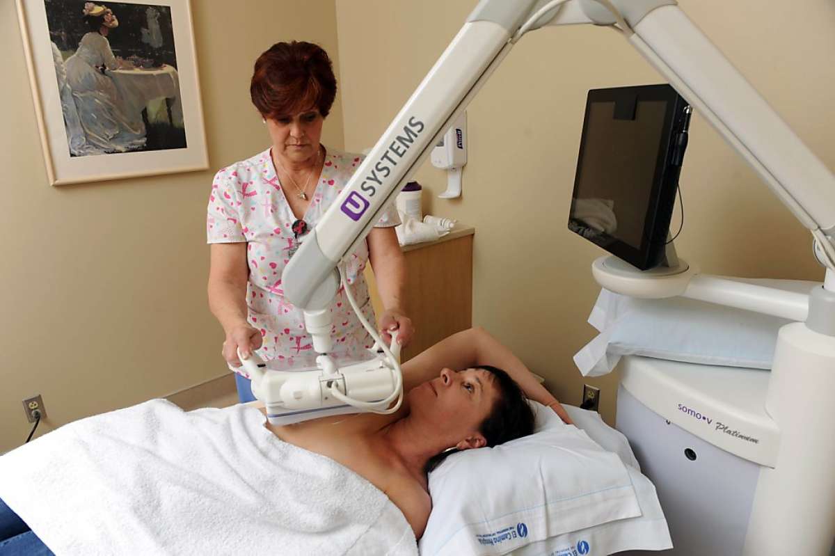
What Is Breast Cancer Screening
Screening exams find disease before symptoms begin. The goal of screening is to detect disease at its earliest and most treatable stage. In order to be widely accepted and recommended by medical practitioners, a screening program must meet a number of criteria, including reducing the number of deaths from the given disease.
Screening tests may include lab tests that check blood and other fluids, genetic tests that look for inherited genetic markers linked to disease, and imaging exams that produce pictures of the inside of the body. These tests are typically available to the general population. However, an individual’s needs for a specific screening test are based on factors such as age, gender, and family history.
In breast cancer screening, a woman who has no signs or symptoms of breast cancer undergoes a breast examination such as:
Bcr/abl Fluorescent In Situ Hybridization
The National Comprehensive Cancer network Myeloproliferative Neoplasms guidelines , state that FISH or a multiplex reverse transcriptase polymerase chain reaction on peripheral blood to detect BCR-ABL1 transcripts and exclude the diagnosis of CML is especially recommended for patients with left-shifted leukocytosis and/or thrombocytosis with basophilia.
Association Of Breast Cancer Screening Behaviors With Stage At Breast Cancer Diagnosis And Potential For Additive Multi
- 1Thrive, An Exact Sciences Company, Cambridge, MA, United States
- 2Blue Health Intelligence, Chicago, IL, United States
- 3Deverka Consulting, LLC, Apex, NC, United States
Purpose: To evaluate mammography uptake and subsequent breast cancer diagnoses, as well as the prospect of additive cancer detection via a liquid biopsy multi-cancer early detection screening test during a routine preventive care exam .
Methods: Patients with incident breast cancer were identified from five years of longitudinal Blue Health Intelligence® claims data and their screening mammogram and PCE utilization were characterized. Ordinal logistic regression analyses were performed to identify the association of a biennial screening mammogram with stage at diagnosis. Additional screening opportunities for breast cancer during a PCE within two years before diagnosis were identified, and the method extrapolated to all cancers, including those without recommended screening modalities.
The study used claims data to demonstrate the association of cancer screening with cancer stage at diagnosis and demonstrates the unmet potential for a MCED screening test which could be ordered during a PCE.
Don’t Miss: What Is Er+ Breast Cancer
What Happens If Something Is Detected On My Screening Exam
Lumps, other abnormalities or questionable findings in the breast are often detected by screening tests. However, it is not always possible to tell from these imaging tests whether a finding is benign or cancerous. To determine whether there is a cancer present, your doctor may recommend that one or more of the following imaging tests may be performed:
- diagnostic mammography
- breast ultrasound
- breast MRI
If a finding is proven to be benign by its appearance on these exams, no further steps may need to be taken. If these tests do not clearly show that the finding is benign, a biopsy may be necessary. In a biopsy, a small amount of tissue is removed under local anesthesia so that it can be examined in a laboratory. One of the following image-guided procedures is used during a breast biopsy:
A pathologist examines the removed tissue specimen and makes a final diagnosis. Depending on the facility, the radiologist or your referring physician will share the results with you.
With early detection and improved treatments, more women are surviving breast cancer. If cancer is diagnosed, your doctor will discuss your treatment options and together you will determine your course of treatment. Today, women have more treatment options than ever before. For more information on treatment, see the Breast Cancer Treatment page.
What Are Dense Breasts

It is very common for women to be told that they have dense breasts after a mammogram. Dense breasts are completely normal and tend to be more common in younger women and in women with smaller breasts. But anyone regardless of age or breast size can have dense breasts.
A doctor will tell you that your breasts are dense if most of the tissue seen on your mammogram is fibrous or glandular breast tissue. These tissue types appear thicker and denser than fatty tissue and will show up white on a mammogram. Because cancer cells also appear white on the image, it may be harder for radiologists to identify disease in women with dense breasts. So thats why some women with dense breasts may be asked to undergo additional imaging tests, such as ultrasound or MRI, which can pick up some cancers that may be missed on a mammogram.
Recommended Reading: Breast Cancer Stage 3
Could You Have Breast Cancer
Breast cancer is a common type of cancer that starts in the breast. About one in eight women will develop breast cancer at some point in their life, and for every 100,000 women diagnosed with breast cancer, about 20 will die from it. Breast cancer risk increases with age every year, over 80% of cases are diagnosed in women over age 50.
Although breast cancer is more common in women, men can get it too. In fact, about 1 in every 100 breast cancer is diagnosed in a man.
How To Determine Ntrk Fusions Status
There are different methods for identifying NTRK fusions: fluorescence in situ hybridization , RT-PCR, and RNA- or DNA-based NGS. Notably, IHC may be used as a screening method, as we will discuss below .
The use of FISH or RT-PCR is not recommended as a screening tool and should be reserved for cases where NTRK fusions are highly recurrent as in the case of infantile fibrosarcoma or secretory breast carcinoma. FISH can be performed with break-apart probes for the three NTRK genes, which requires either separate or multiplex assays, or through a break-apart probe for the ETV6 gene in cases that are histologically suggestive of ETV6-NTRK3 fusions. FISH is not able to identify the gene fusion partner, requires expertise, and is more expensive when a multiplex assay is used. RT-PCR provides direct evidence of a NTRK fusion and detects only known fusion partners and breakpoints,.
RNA- or DNA-based NGS methods are able to assess NTRK fusions with the advantage of providing other important molecular information, including the presence of other oncogenic drivers, tumor mutation burden, and monitoring of patients for the development of resistance mutations. RNA-based NGS has some advantages over DNA, since it is an approach that allows de novo detection of gene fusion transcripts that have not been previously described and increases the sensitivity of detection in low tumor purity samples.
Also Check: Estrogen Positive Progesterone Negative Breast Cancer
Liquid Biopsy For Lung Cancer
An ASCO and College of American Pathologists joint review of circulating tumor DNA analysis in patients with cancer concluded: “Some ctDNA assays have demonstrated clinical validity and utility with certain types of advanced cancer however, there is insufficient evidence of clinical validity and utility for the majority of ctDNA assays in advanced cancer. Evidence shows discordance between the results of ctDNA assays and genotyping tumor specimens and supports tumor tissue genotyping to confirm undetected results from ctDNA tests. There is no evidence of clinical utility and little evidence of clinical validity of ctDNA assays in early-stage cancer, treatment monitoring, or residual disease detection. There is no evidence of clinical validity and clinical utility to suggest that ctDNA assays are useful for cancer screening, outside of a clinical trial.”
Breast Cancer Screening Health Professional Version
On This Page
Note: Separate PDQ summaries on Breast Cancer Prevention, Breast Cancer Treatment , Male Breast Cancer Treatment, and Breast Cancer Treatment During Pregnancy are also available.
Mammography is the most widely used screening modality for the detection of breast cancer. There is evidence that it decreases breast cancer mortality in women aged 50 to 69 years and that it is associated with harms, including the detection of clinically insignificant cancers that pose no threat to life . The benefit of mammography for women aged 40 to 49 years is uncertain. There are randomized trials in India, Iran, and Egypt that have studied the use of clinical breast examination as a screening test. Some of these studies have suggested a shift in late-stage disease however, there is still insufficient evidence to conclude a mortality benefit. Breast self-exam has been shown to have no mortality benefit.
Technologies such as ultrasound, magnetic resonance imaging, and molecular breast imaging are being evaluated, usually as adjuncts to mammography, and are not primary screening tools in the average population.
Informed medical decision making is increasingly recommended for individuals who are considering cancer screening. Many different types and formats of decision aids have been studied.
Don’t Miss: Honey And Baking Soda Cancer Treatment
If You Have A Family History Of Breast Cancer
UK guidelines recommend that women with a moderate or high risk of breast cancer because of their family history should start having screening mammograms every year in their forties.
If you are younger than 40 and have an increased risk of breast cancer, you should be offered yearly MRI scans from the age of 30 or 40. This depends on your level of risk.
You May Like: How Serious Is Grade 3 Breast Cancer
Measurement Of Circulating Tumor Cells For Screening Of Colorectal Cancer
Lopresti and associates stated that CTCs represent an easy, repeatable and representative access to information regarding solid tumors. However, their detection remains difficult because of their paucity, their short half-life, and the lack of reliable surface biomarkers. Flow cytometry is a fast, sensitive and affordable technique, ideal for rare cells detection. Adapted to CTCs detection , most FC-based techniques require a time-consuming pre-enrichment step, followed by a 2-hours staining procedure, impeding on the efficiency of CTCs detection. These researchers overcame these caveats and reduced the procedure to less than 1 hour, with minimal manipulation. First, cells were simultaneously fixed, permeabilized, then stained. Second, using low-speed FC acquisition conditions and 2 discriminators , these investigators suppressed the pre-enrichment step. Applied to blood from donors with or without known malignant diseases, this protocol ensured a high recovery of the cells of interest independently of their epithelial-mesenchymal plasticity and could predict which samples were derived from cancer donors. The authors concluded that this proof-of-concept study laid the bases of a sensitive tool to detect CTCs from a small amount of blood upstream of in-depth analyses .
Furthermore, National Comprehensive Cancer Networks clinical practice guidelines on “Colon cancer” and “Rectal cancer” do not mention measurement of circulating tumor cells as a screening tool.
Also Check: Symptoms Of Ductal Breast Cancer
When And How Often To Get Screened
The decision of when and how often to have breast cancer screening is up to you, based on discussions with your doctor about your personal risks for breast cancer and the benefits of screening.
Breast cancer screening guidelines are generally divided into two groups:
- Guidelines for women at average risk of getting breast cancer
- Guidelines for women at high risk of developing breast cancer
Breast cancer risk is determined based on a combination of your risk factors for breast cancer and other data sets or models, but it is only an estimate. It can vary depending on the assessment tools used.
Mismatch Repair Deficiency And High Microsatellite Instability

Microsatellites are repeated sequences of 16 nucleotides and are mostly located near the ends of chromosomes. They are particularly susceptible to acquired errors when the mismatch repair system is impaired. The MMR system represents one of the DNA repair pathways and corrects DNA base substitution mismatch, insertion or deletion, and slippageconditions generated by DNA replication errors. MMR deficiency arises due to mutations in at least one of the genes that encodes proteins in the MMR system or through methylation of the MLH1 gene promoter that leads to MSI through accumulations of errors in DNA microsatellites.
Recommended Reading: Stage 4 Breast Cancer Prognosis Without Treatment
Oncosignal Test For Analysis Of Solid Tumors
According to Protean BioDiagnostics, OncoSignal is a new way to analyze breast cancer and other cancers. The OncoSignal test uses an advanced molecular and bioinformatics system to measure mRNA expression patterns and calculate the specific activity of 7 key oncogenic driver signal pathways, which include ER , AR , PI3K , HH , NOTCH , TGFbeta , and MAPK . The pathways measure key oncogenic drivers of numerous distinct cancer types including but not limited to breast cancer, prostate cancer, ovarian cancer, colon cancer, lymphoma and more.
Quantifying The Opportunity For An Additive Blood
In order to quantify the target opportunity of an additive blood-based screening test ordered during a PCE, we evaluated both current breast cancer screening rates and potential screening rates if there were widespread adoption of such a test. For breast cancer, the potential screening rate was the proportion of breast cancer patients who had either a screening mammogram or PCE in the two years before their diagnosis. For other cancer types, we looked at tumor types which make up the top 5 leading causes of cancer death without USPSTF-recommended screening modalities available. We limited this list to solid tumors, which are the focus of blood-based multi-cancer screening tests currently in development . Cancer types that meet these criteria include prostate, pancreatic, liver, lymphoma, and ovarian cancers . The potential screening rate for these cancers was defined as the proportion of patients with these tumor types that had a PCE in the two years before their diagnosis date. Colorectal, lung, and cervical cancers were not included in this analysis as their current screening rates were not studied with these data.
You May Like: Does Cancer Hurt In Breast
Neurotrophic Tropomyosin Receptor Kinase Fusions
The NTRK1, NTRK2, and NTRK3 genes encode the neurotrophin receptors TRKA, TRKB, and TRBC, respectively, which are predominantly transcribed in the nervous system in adult tissues. The TRK family plays an important role in nervous system development through regulation of cell proliferation, differentiation, apoptosis, and survival of neurons in both central and peripheral nervous systems.
Fusions involving these genes are the most common mechanisms of oncogenic TRK activation and are found in both adult and pediatric tumors. NTRK fusions are enriched in rare cancer types, including infantile fibrosarcoma, congenital mesoblastic nephroma, secretory breast carcinoma, and mammary analog secretory carcinoma. Common tumors, such as lung, melanoma, and colorectal cancers, have low frequencies of these genomic alterations.
Understanding Abnormal Cervical Screening Test Results
Your current screening test results along with your past test results, determine your risk of developing cervical cancer. Your doctor will use them to figure out your next test or treatment. It could be a follow-up screening test in a year, a colposcopy, or one of the other procedures discussed below to treat any pre-cancers that might be found.
Because there are many different follow-up or treatment options depending on your specific risk of developing cervical cancer, it is best to talk to your healthcare provider about your screening results in more detail, to fully understand your risk of cervical cancer and what follow-up plan is best for you.
Also Check: Breast Cancer Life Expectancy Without Treatment
If You Have A Normal Result
You will receive a letter to let you know your mammogram does not show any signs of cancer. Your next screening appointment will be in 3 years time. Do contact your GP or local screening unit if you havent received an appointment and think you are due one.
It is important to see your GP If you notice any symptoms between your screening mammograms.
You May Like: Why Is Left Breast Cancer More Common
How Often Should I Do Breast Cancer Screening Tests
For women at average risk:
-
Between 40 and 49 years old, starting at age 40, women can consider getting a mammogram every year. Some organizations recommend getting them every two years.
-
Between 40 and 74 years old, women should be screened every 1 to 2 years.
For women at high risk:
-
Starting by age 30 , women should get an annual mammogram and MRI. If an MRI is not an option, an ultrasound can be considered.
-
For women with a history of chest radiation, an annual mammogram and MRI are recommended starting 8 years after radiation treatment .
Women at high risk of getting breast cancer should be screened regularly from a young age, and consider having a breast MRI in addition to a yearly mammogram especially before menopause. The guidelines arent as clear as to when screening should stop. Some organizations recommend that screening should continue until a woman is expected to only live for 5 to 7 more years based on her health or age, or until a woman would no longer want treatment even if she had an abnormal screening test.
Recommended Reading: What Does Stage 3 Cancer Mean
Nf1 Ret And Sdhb For Ovarian Cancer
Norris and stated that neurofibromatosis type 1 is caused by mutations in the NF1 gene encoding neurofibromin, which negatively regulates Ras signaling. NF1 patients have an increased risk of developing early onset breast cancer, however, the association between NF1 and high grade serous ovarian cancer is unclear. Since most NF1-related tumors exhibit early bi-allelic inactivation of NF1, the authors evaluated the evolution of genetic alterations in HGSOC in an NF1 patient. Somatic variation analysis of WES of tumor samples from both ovaries and a peritoneal metastasis showed a clonal lineage originating from an ancestral clone within the left adnexa, which exhibited copy number loss of heterozygosity in the region of chromosome 17 containing TP53, NF1, and BRCA1 and mutation of the other TP53 allele. This event led to bi-allelic inactivation of NF1 and TP53 and LOH for the BRCA1 germline mutation. Subsequent CN alterations were found in the dominant tumor clone in the left ovary and nearly 100 % of tumor at other sites. Neurofibromin modeling studies suggested that the germline NF1 mutation could potentially alter protein function. The authors concluded that these findings demonstrated early, bi-allelic inactivation of neurofibromin in HGSOC and highlighted the potential of targeting RAS signaling in NF1 patient.