Stage 1 Breast Cancer Symptoms
- Hormone therapy
Some treatments are used in a specific order to either shrink a tumor before surgery or to minimize the risk of cancer recurrence after surgery. Treatments given before surgery are called neoadjuvant therapies, while post-surgical treatments are called adjuvant therapies.
Stage 1 breast cancer surgery typically includes removing and testing one or more lymph nodes to see whether the cancer has spread.
Genomic Tests To Predict Recurrence Risk
Doctors use genomic tests, also called multigene panels, to test a tumor to look for specific genes or proteins that are found in or on cancer cells. These tests help doctors better understand the unique features of a person’s breast cancer. Genomic tests can also help estimate the risk of the cancer coming back after treatment. Knowing this information helps doctors and patients make decisions about specific treatments and can help some patients avoid unwanted side effects from a treatment they may not need.
Genomic tests are different from genetic tests. Genetic tests are performed on blood or saliva and are used to determine what gene changes a person may have inherited from a parent that may increase their risk of developing breast cancer. The results of a few genetic tests can also be used to make decisions about specific treatments.
The genomic tests listed below can be done on a sample of the tumor that was already removed during biopsy or surgery. Most patients will not need an extra biopsy or more surgery for these tests.
For patients age 50 or younger who have cancer in 1 to 3 lymph nodes
-
Recurrence score less than 26: Chemotherapy is often recommended before hormonal therapy is given
-
Recurrence score of 26 or higher: Chemotherapy is usually recommended before hormonal therapy is given
For patients older than 50 who do not have cancer in any lymph nodes or who have cancer in 1 to 3 lymph nodes
How Are Breast Ultrasound Results Reported
Doctors use the same standard system to describe results of mammograms, breast ultrasound, and breast MRI. This system sorts the results into categories numbered 0 through 6.
For more details on the BI-RADS categories, see Understanding Your Mammogram Report. While the categories are the same for each of these imaging tests, the recommended next steps after these tests might be different.
You May Like: How To Know If You Have Breast Cancer Gene
Signs And Symptoms Of Inflammatory Breast Cancer
Inflammatory breast cancer causes a number of signs and symptoms, most of which develop quickly , including:
- Swelling of the skin of the breast
- Redness involving more than one-third of the breast
- Pitting or thickening of the skin of the breast so that it may look and feel like an orange peel
- A retracted or inverted nipple
- One breast looking larger than the other because of swelling
- One breast feeling warmer and heavier than the other
- A breast that may be tender, painful or itchy
- Swelling of the lymph nodes under the arms or near the collarbone
If you have any of these symptoms, it does not mean that you have IBC, but you should see a doctor right away. Tenderness, redness, warmth, and itching are also common symptoms of a breast infection or inflammation, such as mastitis if youre pregnant or breastfeeding. Because these problems are much more common than IBC, your doctor might suspect infection at first as a cause and treat you with antibiotics.
Treatment with antibiotics may be a good first step, but if your symptoms dont get better in 7 to 10 days, more tests need to be done to look for cancer. Let your doctor know if it doesn’t help, especially if the symptoms get worse or the affected area gets larger. The possibility of IBC should be considered more strongly if you have these symptoms and are not pregnant or breastfeeding, or have been through menopause. Ask to see a specialist if youre concerned.
How Do I Get Ready For A Breast Ultrasound

-
Your healthcare provider will explain the procedure to you. Ask any questions you have about the procedure.
-
You may be asked to sign a consent form that gives permission to do the test. Read the form carefully and ask questions if anything is not clear.
-
You do not need to stop eating or drinking before the test. You also will not need medicine to help you relax.
-
You should not put any lotion, powder, or other substances on your breasts on the day of the test.
-
Wear clothing that you can easily take off. Or wear clothing that lets the radiologist or technologist reach your chest. The gel put on your skin during the test does not stain clothing, but you may want to wear older clothing. The gel may not be completely removed from your skin afterward.
-
Follow any other instructions your healthcare provider gives you to get ready.
Also Check: Stage 2 Breast Cancer Survival Rate After 20 Years
What Are Screening Tests
Screening refers to tests and exams used to find a disease in people who dont have any symptoms. The goal of screening tests for breast cancer is to find it early, before it causes symptoms . Early detection means finding and diagnosing a disease earlier than if youd waited for symptoms to start.
Breast cancers found during screening exams are more likely to be smaller and less likely to have spread outside the breast. The size of a breast cancer and how far it has spread are some of the most important factors in predicting the prognosis of a woman with this disease.
What Is An Ultrasound
Ultrasound screening is commonly used to study a developing fetus, heart and blood vessels, muscles, and tendons, and abdominal and pelvic organs. Beyond that, your radiologist would recommend an ultrasound scan when you get an abnormal mammogram.
The ultrasound uses high-frequency sound waves to look for tumors in the breast when they dont show up on X-rays .
In a breast ultrasound scan, your healthcare provider moves a wand-like device called a transducer over your breasts to get their images.
The transducer sends high-frequency sound waves that bounce off your breast tissue. It then picks up the bounced sound waves to develop pictures of the inside of your breasts.
Don’t Miss: Can Breast Cancer Cause Fluid In The Lungs
American Cancer Society Screening Recommendations For Women At Average Breast Cancer Risk
The COVID-19 pandemic initially resulted in most elective procedures being put on hold, leading to many people not getting screened for cancer. Learn how you can talk to your doctor and what steps you can take to plan, schedule, and get your regular cancer screenings in Cancer Screening & COVID-19.
These guidelines are for women at average risk for breast cancer. For screening purposes, a woman is considered to be at average risk if she doesnt have a personal history of breast cancer, a strong family history of breast cancer, or a genetic mutation known to increase risk of breast cancer , and has not had chest radiation therapy before the age of 30.
- Women between 40 and 44 have the option to start screening with a mammogram every year.
- Women 45 to 54 should get mammograms every year.
- Women 55 and older can switch to a mammogram every other year, or they can choose to continue yearly mammograms. Screening should continue as long as a woman is in good health and is expected to live at least 10 more years.
- All women should understand what to expect when getting a mammogram for breast cancer screening what the test can and cannot do.
Clinical breast exams are not recommended for breast cancer screening among average-risk women at any age.
Vegfr2 Expression Increases With Breast Tissue Progression From Normal Hyperplasia Dcis To Invasive Breast Cancer
Representative photomicrographs of mouse mammary glands with four different histological stages of breast cancer development. Mammary gland sections were stained for vascular endothelial cell marker CD31 and for VEGFR2 . Merged images show expression of VEGFR2 on vascular endothelial cells. Note that number of microvessels and the VEGFR2 staining increased with breast cancer development .
Also Check: Mortality Rates Of Breast Cancer
What Is Breast Ultrasound
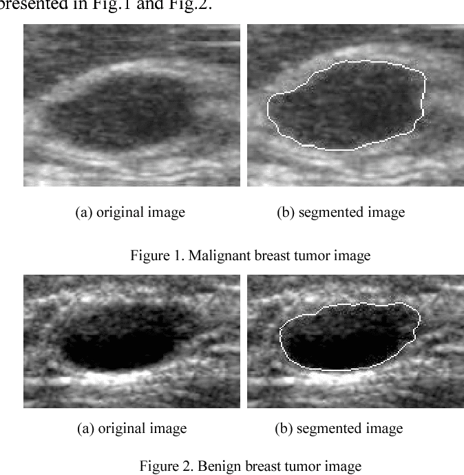
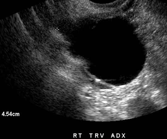
Breast ultrasound is an imaging test that uses sound waves to look at the inside of your breasts. It can help your healthcare provider find breast problems. It also lets your healthcare provider see how well blood is flowing to areas in your breasts. This test is often used when a change has been seen on a mammogram or when a change is felt, but does not show up on a mammogram.
The healthcare provider moves a wand-like device called a transducer over your skin to make the images of your breasts. The transducer sends out sound waves that bounce off your breast tissue. The sound waves are too high-pitched for you to hear. The transducer then picks up the bounced sound waves. These are made into pictures of the inside of your breasts.
Your healthcare provider can add another device called a Doppler probe to the transducer. This probe lets your healthcare provider hear the sound waves the transducer sends out. He or she can hear how fast blood is flowing through a blood vessel and in which direction it is flowing. No sound or a faint sound may mean that you have a blockage in the flow.
Ultrasound is safe to have during pregnancy because it does not use radiation. It is also safe for people who are allergic to contrast dye because it does not use dye.
How To Determine The Grade Of Breast Cancer
There are different scoring systems available for determining the grade of a breast cancer. One of these systems is the Nottingham Histologic Score system . In this scoring system, there are three factors that the pathologists take into consideration: 1 the amount of gland formation 2 the nuclear features 3 the mitotic activity
Don’t Miss: Warning Sign Of Breast Cancer
Stage 0 Breast Cancer Symptoms
Very early-stage DCIS breast cancers typically dont have symptoms. Though its sometimes possible to feel a small, hard lump, most people discover they have stage 0 breast cancer through regular mammogram screenings.
Pagets disease of the breast is likely to cause noticeable changes, such as:
- Red, crusty or scaly skin of the nipple or areola
- Yellow fluid discharge from the nipple
- Other nipple discharge
- A flat or inverted nipple
- Burning or itching of the breast or nipple
Assessment Categories For Breast Ultrasound

The positive predictive value of detecting carcinomas by breast ultrasound mainly derives from the sonographic assessment criteria and categories applied. Overall, the range is still low relative to mammography.
Kaplan, 2001 used a two-armed categorization approach, namely a simple subdivision into negative and positive. All positive results were considered to be potentially suspicious. In contrast to the other studies, Kaplan also deemed cysts positive if they exceeded a size of 1 cm. The author had a low positive predictive value of 2% . Three studies subdivided their findings into three categories, but applied different definitions of these categories. Categories 1, 2 and 3 were called ‘normal’, ‘benign’, and ‘suspicious’ or ‘benign’, ‘indeterminate’ and ‘malignant’ . These authors found positive predictive values of biopsy of 10.3% and 28% . The values for Leconte’s study could not be ascertained . Crystal et al., 2003 used four arms of classification, thereby reaching a positive predictive value for malignancy of 20%. Other than Buchberger et al., Crystal et al. included the ‘indeterminate’ findings in their calculation of suspicious findings. Only one study used the five categories proscribed by the BI-RADS classification for breast ultrasound scans . In this study, however, the positive predictive value could not be determined.
Recommended Reading: Can You Work During Breast Cancer Treatment
When Is Breast Ultrasound Used
Ultrasound is not typically used as a routine screening test for breast cancer. But it can be useful for looking at some breast changes, such as lumps . Ultrasound can be especially helpful in women with dense breast tissue, which can make it hard to see abnormal areas on mammograms. It also can be used to get a better look at a suspicious area that was seen on a mammogram.
Ultrasound is useful because it can often tell the difference between fluid-filled masses like cysts and solid masses .
Ultrasound can also be used to help guide a biopsy needle into an area of the breast so that cells can be taken out and tested for cancer. This can also be done in swollen lymph nodes under the arm.
Ultrasound is widely available and is fairly easy to have done, and it does not expose a person to radiation. It also tends to cost less than other testing options.
Skin Rash On The Breasts
You may not associate breast cancer with redness or a skin rash, but in the case of inflammatory breast cancer , a rash is an early symptom. This is an aggressive form of breast cancer that affects the skin and lymph vessels of the breast.
Unlike other types of breast cancer, IBC doesnt usually cause lumps. However, your breasts may become swollen, warm, and appear red. The rash may resemble clusters of insect bites, and its not unusual to have itchiness.
Don’t Miss: Can Breast Cancer Lead To Lung Cancer
Prediction Of Aln Status Between N0 And N+
Adopting N0 as negative reference standard, 466 lesions were randomly assigned as training cohort and the other 118 lesions as independent test cohort. The detailed characteristics including patient age, US size, Breast Imaging-Reporting and Data System category, tumor type, estrogen receptor status, progesterone receptor status, human epidermal growth factor receptor 2 , Ki-67 proliferation index were demonstrated in Table . There was no significant difference between the detailed characteristics of the two cohorts . Based on axillary US findings evaluated by an experienced radiologist, axillary US findings had an AUC of 0.735, accuracy of 0.635, sensitivity of 0.721 and specificity of 0.573. The Kappa values for axillary US were 0.933 for inter-observer agreement and 1 for intra-observer agreement .
Table 2 Patient and tumor characteristics.Table 3 The prediction of ALN status results ).Fig. 2: Comparison of receiver operating characteristic curves between different models for predicting disease-free axilla and any axillary metastasis ).
DLR deep learning radiomics. Numbers in parentheses are areas under the receiver operating characteristic curves. Source data are provided as a Source Data file.
Changes In The Size And Shape Of The Breast
Its not uncommon for breasts to swell, and you may notice a change in size around the time of your menstrual cycle.
Swelling can also cause breast tenderness, and it may be slightly uncomfortable to wear a bra or lie down on your stomach. This is perfectly normal and rarely indicative of breast cancer.
But while your breasts may undergo certain changes at different times of the month, you shouldnt overlook some changes. If you notice your breasts swelling at times other than your menstrual cycle, or if only one breast is swollen, talk to your doctor.
In cases of normal swelling, both breasts remain symmetrical. That means one wont suddenly be larger or more swollen than the other.
Read Also: Alternative Treatments For Breast Cancer
Cancer Diagnosis According To Breast Density Categories
Two studies analyzed mammographic results of breasts in categories ACR 3 to ACR 4 , and the other studies evaluated the mammograms of ACR 2 to ACR 4 breast tissue .
Women with breasts of types ACR 3 or ACR 4 proved to have the highest proportion of breast cancers diagnosed by ultrasound screening.
Leconte et al. diagnosed 16 carcinomas in all 11 of which were detected in ACR 3 and ACR 4 breast tissue and five in women with breasts in categories ACR 1 and 2. Buchberger et al. diagnosed 36 malignancies in breast tissue of ACR types 3 and 4 , while two carcinomas were found in the ACR category 2 breasts . Crystal et al. found no carcinomas in ACR-2 women, and 0.4% and 0.3% in breast-density categories ACR 3 and ACR 4, respectively. Kaplan diagnosed cancer at the rate of 0.11% in ACR 2 breast tissue and 0.27% and 0.25% in ACR categories 3 and 4, in the one series where technologists performed the screening ultrasound.
Group Of Women Examined
The systematic search yielded studies in which breast ultrasound was used as a supplemental examination following mammographic interpretation. Moreover, of the group of asymptomatic women with negative mammographic results, women with breast tissue density went on to be examined by ultrasound. The only exception is the study performed by Leconte et al., 2003 , where 3% had palpable findings .
The fraction of these women relative to all women screened within the specified period was reported in two studies at 36% and 35.8% . The size of the study populations ranged from n = 1517 to n = 13547, with mean n = 5118.
Also Check: Can You Get Breast Cancer From Sleeping On Your Stomach
Criteria For Inclusion And Exclusion
In order to ensure evaluability and comparability, the studies were required to contain the following information to be included in the present analysis:
– breast density according to the BI-RADS ACR categories and/or quantification ACR 1: almost entirely fat , ACR 2: scattered fibroglandular densities , ACR 3: heterogeneously dense , ACR 4: extremely dense .
For inclusion, all of the following criteria were required to be satisfied:
1. study/review deals with the questions under investigation
2. adequate type of study
3. adequate study population
4. intervention complies with technical standards
5. required data are provided
Publications were excluded if any of the following criteria were met:
1. redundancy: multiple publications of the same data
2. methods: case reports, expert opinions, or poor-quality case-control studies
What Are The Side Effects Of Breast Cancer

Complications from this cancer type include the spread of cancer from the breast to other locations and treatment side effects that may include nausea, vomiting, and hair loss. In some cases, the tumor is in an advanced stage of progression this can result in severe signs and symptoms and complications.
Don’t Miss: How To Decrease Risk Of Breast Cancer