Palpation Of Benign Breast Masses
In contrast to breast cancer tumors, benign lumps are often squishy or feel like a soft rubber ball with well-defined margins. They’re often easy to move around and may be tender.
Breast infections can cause redness and swelling. Sometimes it can be difficult to tell the difference between mastitis and inflammatory breast cancer, but mastitis often causes symptoms of fever, chills, and body aches, and those symptoms aren’t associated with cancer.
Ultrasound And Breast Density
Specialists will often use ultrasound to screen women with high breast density because mammograms can be difficult to interpret in this group.
For this reason, ultrasound is frequently a first diagnostic imaging method for women under 35.
Whether or not an ultrasound can stand-alone as a screening method versus combining it with mammography or MRI is still a subject of debate.
At present, there is no study which definitively proves that ultrasound screening alone lowersmortality rates for breast cancer, unlike mammography, which does.
In the past, a woman with dense breast tissue and no abnormalities seen on mammogram would not go on to have an ultrasound.
However, more and more often, when mammograms have very dense tissue, medics will recommend an ultrasound too.
How Long Does A Breast Ultrasound Take
The examination takes between 1530;minutes.
Sometimes you will be asked to wait and have the images checked by a radiologist . Sometimes it will be necessary for the radiologist to attend the examination, because it may be important to see the images on the screen rather than as still photographs. The radiologist may also want to examine your breast if you have a symptom and might also ask you some questions about these symptoms. This extra information may help the radiologist to better understand your ultrasound images, so they can give an accurate diagnosis.
Recommended Reading: What Does Breast Cancer Look Like On The Outside
Combining Ultrasound For Breast Cancer With Mri Or Biopsy
Research shows that the combination of ultrasound with Magnetic Resonance Imaging is a particularly good combination in follow-up evaluation of lesions found on mammography.
The detail of the MRI greatly assists in diagnostic and treatment decisions. Ultrasound is also very useful in guiding the needle during a follow-up biopsy.
Palpation Of Cancerous Masses

Cancerous masses in the breast are often very firm, like a rock or a carrot, and have an irregular shape and size. They are often fixedthey feel like they are attached to the skin or nearby tissue so that you can’t move them around by pushing on thembut can be mobile. They’re also not likely to be painful, though they can be in some cases.
On exam, other changes may be present as well, such as dimpling of the skin or an orange-peel appearance, nipple retraction, or enlarged lymph nodes in the armpit.
One type of breast cancer, inflammatory breast cancer, does not usually cause a lump but instead involves redness, swelling, and sometimes a rash on the skin of the breast.
You May Like: What Does Pt1c Mean In Breast Cancer
What Is It Like Having The Test
Ultrasound can be done in a doctors office, clinic, or hospital. Wear comfortable clothes. Depending on the body part to be studies, you may need to change into a hospital gown.
Most often you will lie down on a table. Your position will depend on the body part to be studied. The technologist will put a water-based gel on your skin and move the transducer over the area to be checked. The gel both lubricates the skin and helps conduct the sound waves. The gel feels cool and slippery. If a probe is used, it will be covered with gel and put into the body opening. This can cause pressure or discomfort.
During the test the technologist or the doctor moves the transducer as its firmly pressed to your skin. You may be asked to hold your breath during the scan. The operator may adjust knobs or dials to increase the depth to which the sound waves are sent. You may feel slight pressure from the transducer.
After the test the gel is wiped off your skin. It does not stain your skin or your clothing.
Mammogram And Ultrasound Images Explained
A mammogram is a routine part of a breast cancer screening program.;;
Physicians agree that breast palpation programs are generally insufficient for early breast cancer detection.
Breast self-examination programs are also unreliable as a lesion can develop for years before it becomes palpable.
Of course, when a family physician finds a bump or a lump of some kind on a clinical exam, he/she will immediately refer the patient for a mammogram. Women with higher than average risk factors and older women should generally have mammograms more frequently.
Normally, the X-ray component of a mammogram is all that is necessary for breast cancer screening purposes.
So, an ultrasound is typically a second look type of application. However, it is not a good idea to have an ultrasound instead of a mammogram and it is probably best to follow the advice of the screening physicians.
I just want to let you know that I have a newer version of this page, with more up-to-date information on Mammogram Screening Images. However, it isnt nearly as long as this one. This page is still really useful.
Don’t Miss: What Is Er Pr Positive Breast Cancer
Evaluation Of A Palpable Mass In A Patient With Negative Mammogram
Fifty years ago, women who presented with a palpable mass eventually underwent surgical excision to exclude malignancy . With advances in ultrasound imaging, many women now who present with a palpable mass and no mammographic correlate undergo diagnostic targeted ultrasound, often on the same day as diagnostic mammogram, to evaluate the region of palpable concern. If no mammographic or sonographic abnormality is identified, women can be safely reassured that there is no abnormality instead of undergoing unnecessary surgery or biopsy . However, if a patient presents with a palpable mass with negative mammogram, ultrasound has been shown to be effective in identifying an abnormality in about 50% of cases, with the majority of these abnormalities characterized as benign or likely benign . Recent studies also question whether a repeat mammogram is even necessary when a woman presents with a new palpable mass within 12 months of prior negative mammogram, given that ultrasound has been shown to yield the most diagnostic information .
Ultrasound Vs Fast Mri For People With Dense Breasts
That said, recent studies suggest that for women who have dense breasts, the combination of mammography and fast breast MRI may be more sensitive and produce fewer false positives than the combination of mammography and ultrasound. Fast breast MRI appears to be relatively comparable to conventional MRI , but takes only around 10 minutes to perform with a cost similar to that of mammography. Since the testing is relatively new, however, it is not currently available at every center that performs breast cancer screening.
Recommended Reading: Can You Get Breast Cancer At 20
Interpreting Breast Cancer Screening Mammograms Improves With Experience
It takes years of radiological experience to gain experience and knowledge in interpreting mammograms. However, anything abnormal, and especially features which show unusual density, odd shapes, and irregular borders, will need a;biopsy.
Interpretation accuracy improves over the first three years of practice and continues to be refined over the course of a radiologists career. For some reason, the rate of abnormal findings on mammograms is slightly higher in North America than in Europe.
Breast Ultrasound For Women At Higher Risk Of Breast Cancer
For women at higher than average;risk of breast cancer, screening with breast ultrasound doesnt appear to add extra benefit to screening with breast MRI and mammography .
Studies are looking at whether breast ultrasound may be a useful addition to screening mammography among women at higher risk of breast cancer for whom breast MRI is not recommended .
Women at higher risk who are recommended breast MRI as part of breast cancer screening, but cant have one for medical reasons, may consider breast ultrasound .
Learn about breast cancer screening recommendations for women at higher than average risk.;
Also Check: How To Help Breast Cancer Awareness
Schedule An Appointment With Us
The effectiveness of a breast ultrasound depends on the skill of the technologist. Any human error might lead to a misinterpretation of the results or overlooked lumps. For this reason, its imperative that you seek ultrasound services from a trusted imaging and mammography center.
The Womens Center at Colorado Springs Imaging and Envision Imaging offer highly reliable ultrasound services with fast turnaround times. Our competent technologists have vast experience in detecting and interpreting imaging results. Get in touch with us;today to schedule your appointment.
Information About The Lesion As Seen On Mammograms And Ultrasounds

On a mammogram, a lesion will usually appear brighter;than the surrounding tissue. This is because things that are denser than fat will stop more x-ray photons, hence they appear brighter.
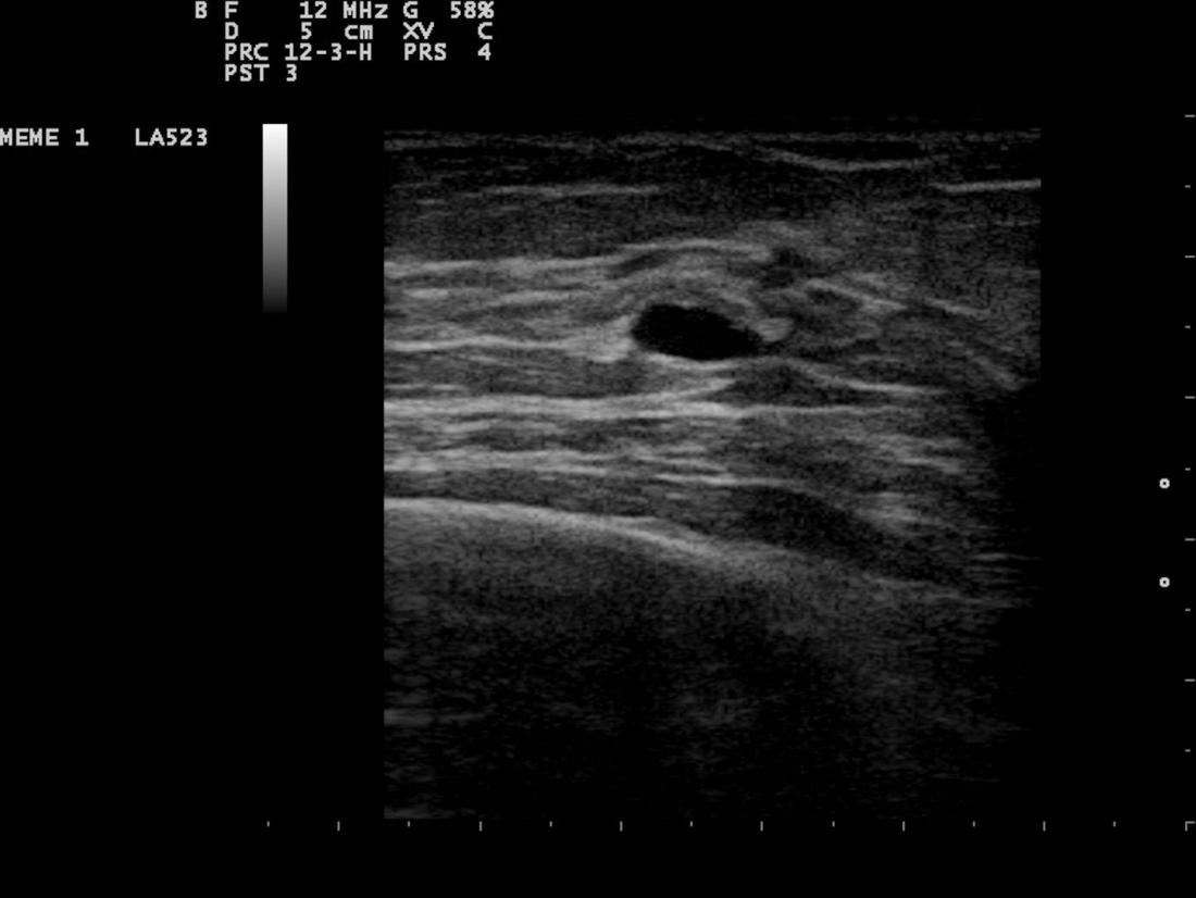
Ultrasounds are a little harder to figure out. The darkest images on a sonograph are cysts containing liquid. Solids are less definitive. With ultrasound, the radiologist will probably be trying to get a sense of the internal texture of the breast lesion and surrounding area.
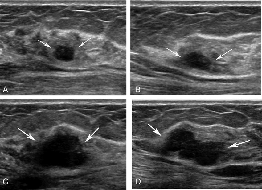
Solid lesions can be a little brighter or darker than the surrounding tissue, and the way to evaluate these on ultrasound is to look closely at the margins or the outer edges;of the nodule.
Also Check: Is Stage 2 Breast Cancer Bad
Chronic Abscess Of The Breast
Patients may present with fever, pain, tenderness to touch and increased white cell count. Abscesses are most commonly located in the central or subareolar area. An abscess may show an ill-defined or a well-defined outline. It may be anechoic or may reveal low-level internal echoes and posterior enhancement .
What Are The Benefits Of A Breast Ultrasound
Ultrasound examination allows the detection and identification of most breast lumps. It is especially useful in distinguishing between solid and fluid-filled lumps.
If the ultrasound does not identify a lump that you or your doctor can feel, then other tests, such as mammography or magnetic resonance imaging , may be required to examine the breast.
Read Also: Can You Get Breast Cancer At 16
Who Interprets The Results And How Do I Get Them
A radiologist, a doctor trained to supervise and interpret radiology exams, will analyze the images. The radiologist will send a signed report to the doctor who requested the exam. Your doctor will then share the results with you. In some cases, the radiologist may discuss results with you after the exam.
You may need a follow-up exam. If so, your doctor will explain why. Sometimes a follow-up exam further evaluates a potential issue with more views or a special imaging technique. It may also see if there has been any change in an issue over time. Follow-up exams are often the best way to see if treatment is working or if a problem needs attention.
How Can This Change My Treatment Plan
If an obviously abnormal node is found before surgery, then you have a more serious cancer. If appropriate, an ultrasound-guided needle biopsy can be performed to confirm the node is involved with cancer. If you have cancer in your nodes, you will likely require chemotherapy either before or after surgery . Regardless of the findings of an axillary ultrasound, a surgical evaluation of your axillary lymph nodes will be needed when you undergo a definitive breast cancer surgery. The surgical procedures used today for lymph nodes are a sentinel node biopsy or an axillary dissection.
Also Check: What Are The Odds Of Breast Cancer Returning
How Long Does An Ultrasound Appointment Take To Come Through
The ultrasound probe is then passed over your skin with light pressure and images of your blood vessels are recorded. You may hear high frequency sounds when we monitor the blood moving in your vessels. The scan can take between 15 to 60 minutes, depending on how much information your doctor has requested.
How Does Tumor Look Like
A tumor is a mass or lump of tissue that may resemble swelling. Not all tumors are cancerous, but it is a good idea to see a doctor if one appears. The National Cancer Institute define a tumor as an abnormal mass of tissue that results when cells divide more than they should or do not die when they should.
You May Like: What Type Of Breast Cancer Is Most Likely To Metastasize
You Feel Like Youve Just Been Punched In The Gut
And you just want to stop for a moment, to take a second to breathe and think…
But you dont have time for that.
You need to make choices about your diagnosis and cancer treatment, now. But these are things that you dont know anything about…and yet will impact your life, forever.
Its not fair…and its terrifying.
Youre smack in the middle of one of the hardest situations anyone can be put in…
You need to make instant decisions that will determine your future and life without knowing anything about whats actually happening in your body and what all of your options are.
How on earth would you know what to do?
And the catch 22, the problem of it all, is that you need to have confidence in your choices if you are to have any peace of mind during this process.
“Alex knows more about cancer than anyone Ive met including doctors. He actually thinks and personalizes treatment for his clients. Doctors are not trained to think and stuck in the past. Highly recommend CTOAM.”
Michael Choe, client
“Alex was just so positive we enjoyed our phone calls with him because we talked to him, we were always like, ‘okay, we can do this!’ There was always hope after we talked to him. There was always something we could be doing more of. You know, drink your green tea, take all your nutraceuticalsthere was just always, always something positive.”
Joan Fleitch, client
Biochemist, Health Educator, and Author Stephen Cherniske of The Healthy Skeptics recommends CTOAM – 5 stars
If You Have An Abnormal Screening Mammogram:

If you have a mammogram, its not uncommon to be called back for an abnormal result. For example, your mammography center might contact you if something is unclear in your mammogram. In the U.S., about 10-12 percent of women are called back after a mammogram for more tests. Its always a good idea to follow up with your doctor about what to do next. The most likely next step is a diagnostic mammogram or breast ultrasound. In some cases, a breast MRI or a biopsy may be recommended.
Here are the different types of follow-up tests:
Mammography can be used as a follow-up test when something abnormal is found on a screening mammogram or CBE. This is called a diagnostic mammogram. The basic procedure for a diagnostic mammogram is the same as for a screening mammogram, but the diagnostic mammogram includes more images of the breast.
Breast ultrasound uses sound waves to make images of the breast. Like a diagnostic mammogram, its considered non-invasive. Breast ultrasound can tell the difference between a liquid-filled cyst and a solid mass .
Breast magnetic resonance imaging uses magnetic fields to create an image of the breast. Its more invasive than mammography because a contrast agent is given through an IV before the test. Breast MRI can be used to get more information after an abnormal mammogram.
Also Check: How Fast Does Breast Cancer Spread To Lymph Nodes
Characterization Of A Mammographic Mass
Ultrasound is an adjunct to mammography for mass characterization and is the next examination to perform for characterization of a mammographic mass, per ACR appropriateness criteria . It is critical to establish the location and depth of the mass identified on mammography to ensure that the same area is imaged during breast ultrasound. If a mass is identified on breast ultrasound and is thought to correlate with the mammographic mass, the size, shape, location, and surrounding tissue composition should correlate between the two modalities . If no sonographic correlate is found for a mass identified on mammogram, then revaluation of the mammogram should be performed. If mammographic findings remain suspicious for a sonographically occult mass, then further evaluation with a different imaging modality and/or biopsy can be pursued .
Figure 16.
. CC mammogram of the left breast and transverse ultrasound of the left breast.
Asymmetric Breast Density Often Has Benign Causes
The X-ray image below shows a lesion with asymmetric density. That indicates that the lesion likely contains a variety of elements, which may or may not indicate breast cancer.;On the sonogram below, the asymmetric density observed in the X-ray appears to be fat tissue. This is due to the fact that although it is a little darker-appearing on ultrasound than other fat, it has an internal texture resembling a fat lobule.
There is an apparent capsule, which is the thin bright line around the outside of the dark oval area. . This suggests the lesion might be hamartoma or fibroadenolipoma, but as there is no apparent capsule on the X-ray, this is less likely.
Usually, hematoma, there would be a collection of solid and liquid components, and that does not appear to be the case here.
The site requires further investigation, perhaps by spot films with compression, .; Likely diagnosis might be dysplasia or other fibrocystic changes.
Don’t Miss: How To Screen For Breast Cancer
Why Would My Doctor Refer Me To Have This Procedure
If you or your doctor can feel a lump in the breast, ultrasound can help to distinguish fluid-filled lumps from solid lumps that may be cancerous or benign .
In younger patients who have breast symptoms , ultrasound is often the first investigation. The breast tissue of younger women is much denser than it is in older women, and this can make it harder to detect an abnormality using an X-ray .
Ultrasound is also used to diagnose problems such as complications from mastitis , assessing abnormal nipple discharge or problems with breast implants.
Ultrasound is commonly used to guide the placement of a needle during biopsies .