Comparison With Other Studies
Owing to the relatively favourable survival rates of patients with breast cancer, long follow-up and large groups of patients are needed to provide sufficient power to detect differences in overall survival. A recent Lancet publication with global data showed age standardised net survival with breast cancer of 80% or more in 34 countries and an increase worldwide but had no data on factors influencing it.1 Our more recent results from 2006-12 even indicated a five year relative survival rate of 96%. Another recent study compared breast cancer recurrence and outcome patterns in 3589 patients treated from 1986 to 1992 matched one to one with patients from 2004-08.3 The authors describe a lower hazard rate of breast cancer relapse and a lower hazard rate of death in the later time period, but similar outcome patterns by oestrogen receptor and HER2 receptor status. Their study was not designed to identify current prognostic factors. This study is difficult to compare with ours, as it differs with respect to the timeframes chosen for the cohorts, the matched design, and the much lower number of patients. Another study by Duffy et al also shows considerably better survival of node negative versus node positive breast cancers in 9040 patients with breast cancer from 1998 to 2003 in the east of England.28
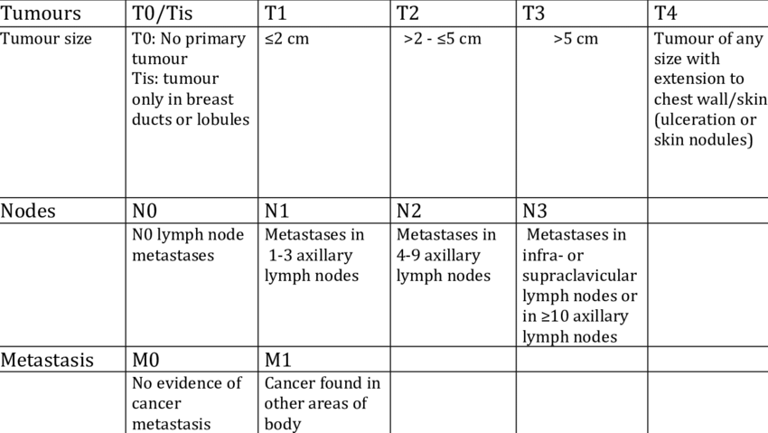
N Categories For Breast Cancer
N followed by a number from 0 to 3 indicates whether the cancer has spread to lymph nodes near the breast and, if so, how many lymph nodes are involved.
Lymph node staging for breast cancer is based on how the nodes look under the microscope, and has changed as technology has gotten better. Newer methods have made it possible to find smaller and smaller groups of cancer cells, but experts haven’t been sure how much these tiny deposits of cancer cells influence outlook.
Its not yet clear how much cancer in the lymph node is needed to see a change in outlook or treatment. This is still being studied, but for now, a deposit of cancer cells must contain at least 200 cells or be at least 0.2 mm across for it to change the N stage. An area of cancer spread that is smaller than 0.2 mm doesn’t change the stage, but is recorded with abbreviations that indicate the type of special test used to find the spread.
If the area of cancer spread is at least 0.2 mm , but still not larger than 2 mm, it is called a micrometastasis . Micrometastases are counted only if there aren’t any larger areas of cancer spread. Areas of cancer spread larger than 2 mm are known to influence outlook and do change the N stage. These larger areas are sometimes called macrometastases, but are more often just called metastases.
NX: Nearby lymph nodes cannot be assessed .
N0: Cancer has not spread to nearby lymph nodes.
N1c: Both N1a and N1b apply.
N3: Any of the following:
N3a: either:
N3b: either:
T Categories For Breast Cancer:
T followed by a number from 0 to 4 describes the main tumor’s size and if it has spread to the skin or to the chest wall under the breast. Higher T numbers mean a larger tumor and/or wider spread to tissues near the breast.
- TX: Primary tumor cannot be assessed
- T0: No sign of primary tumor
- Tis: Carcinoma in situ
Don’t Miss: Side Effects Of Hormone Blockers For Breast Cancer
Independent Predictive Factors In The Training Set
Age, race, and tumour size, grade, primary site, histologic type and subtype were identified as being significantly associated with positive lymph nodes . All significant factors in the univariate analysis were included in the multivariate logistic regression analysis . Histological type was not an independent predictor in our study . All the other variables showed statistically significant predictive capability for positive lymph nodes . The results showed that hormone receptor -/HER2+ patients had more positive lymph nodes compared to HR+/HER2+ patients . There was no significant difference between HR+/HER2- patients and triple negative patients .
What Is Stage 0 Breast Cancer

Also called carcinoma in situ, stage 0 is the earliest breast cancer stage. At stage 0, the breast mass is noninvasive, and there is no indication that the tumor cells have spread to other parts of the breast or other parts of the body. Often, stage 0 is considered a precancerous condition that typically requires close observation, but not treatment.
Stage 0 breast cancer is difficult to detect. There may not be a lump that can be felt during a self-examination, and there may be no other symptoms. However, breast self-exams and routine screening are always important and can often lead to early diagnosis of breast cancer, when the cancer is most treatable. Stage 0 disease is most often found by accident during a breast biopsy for another reason, such as to investigate an unrelated breast lump.
There are two types of stage 0 breast cancer:
Ductal carcinoma in situ occurs when breast cancer cells develop in the breast ducts. Today, stage 0 DCIS is being diagnosed more often because more women are having routine mammogram screenings. DCIS can become invasive, so early treatment can be important.
Don’t Miss: How Curable Is Breast Cancer
Grading Invasive Breast Cancer Cells
Three features of the invasive breast cancer cell are studied and each is given a score. The scores are then added to get a number between 3 and 9 that is used to get a grade of 1, 2, or 3, which is noted on your pathology report. Sometimes the terms well differentiated, moderately differentiated, and poorly differentiated are used to describe the grade instead of numbers:
- Grade 1 or well differentiated . The cells are slower-growing, and look more like normal breast cells.
- Grade 2 or moderately differentiated . The cells are growing at a speed of and look like cells somewhere between grades 1 and 3.
- Grade 3 or poorly differentiated . The cancer cells look very different from normal cells and will probably grow and spread faster.
Our information about pathology reports can help you understand details about your breast cancer.
Changes To The Tnm Classification Systems In The 7th Edition Ajcc Cancer Staging Manual
The newest edition of the American Joint Committee on Cancer, Cancer Staging Manual has only minor changes from the 6th edition.
Within the P or pathology categories, only ductal and lobular carcinoma in situ , and isolated Pagets disease of the nipple are classified as pTis. So-called precursorbreast neoplasm such as atypical ductal or lobular hyperplasia are no longer included.
Some new guidelines are also given reflecting the classification of micro metastasis in the regional lymph nodes. Now, small clusters of cancer cells no larger than 0.2 mm, or non confluent or nearly confluent clusters of cells not exceeding 200 cells in a single histologic lymph node cross section, may be classified as isolated tumor cells ).
And finally, Stage I breast tumors have now been subdivided into Stage IA and Stage IB, with stage IB including small tumors with lymph node micro metastases .
Also Check: What Are The Side Effects Of Breast Cancer
Stages Of Breast Cancer
The stages of breast cancer range from 0 to IV .
The highest stage is any cancer with metastases , no matter the size of the tumor, the lymph node status or other factors. This is known as metastatic breast cancer and is the most advanced stage of breast cancer.
Most often, the higher the stage of the cancer, the poorer the prognosis will be.
The table below lists the TNM classifications for each stage of breast cancer for people who have surgery as their first treatment.
| When TNM is |
| IV |
|
*T1 includes T1mi. **N1 does not include N1mi. T1 N1mi M0 and T0 N1mi M0 cancers are included for prognostic staging with T1 N0 M0 cancers of the same prognostic factor status. ***N1 includes N1mi. T2, T3 and T4 cancers with N1mi are included for prognostic staging with T2 N1, T3 N1 and T4 N1, respectively. |
|
Used with permission of the American College of Surgeons, Chicago, Illinois. The original source for this information is the AJCC Cancer Staging Manual, Eighth Edition published by Springer International Publishing. |
Examples Using The Full Staging System
Because there are so many factors that go into stage grouping for breast cancer, it’s not possible to describe here every combination that might be included in each stage. The many different possible combinations mean that two women who have the same stage of breast cancer might have different factors that make up their stage.
Here are 3 examples of how all of the factors listed above are used to determine the pathologic breast cancer stage:
You May Like: Did Anne Hathaway Have Cancer
How A Breast Cancers Stage Is Determined
Your pathology report will include information that is used to calculate the stage of the breast cancer that is, whether it is limited to one area in the breast, or it has spread to healthy tissues inside the breast or to other parts of the body. Your doctor will begin to determine this during surgery to remove the cancer and look at one or more of the underarm lymph nodes, which is where breast cancer tends to travel first. He or she also may order additional blood tests or imaging tests if there is reason to believe the cancer might have spread beyond the breast.
Breast Cancer Stage Groups
In breast cancer, stage is based on the size and location of the primary tumor, the spread of cancer to nearby lymph nodes or other parts of the body, tumor grade, and whether certain biomarkers are present. To plan the best treatment and understand your prognosis, it is important to know the breast cancer stage.
There are 3 types of breast cancer stage groups:
- Clinical Prognostic Stage is used first to assign a stage for all patients based on health history, physical exam, imaging tests , and biopsies. The Clinical Prognostic Stage is described by the TNM system, tumor grade, and biomarker status . In clinical staging, mammography or ultrasound is used to check the lymph nodes for signs of cancer.
- Pathological Prognostic Stage is then used for patients who have surgery as their first treatment. The Pathological Prognostic Stage is based on all clinical information, biomarker status, and laboratory test results from breast tissue and lymph nodes removed during surgery.
- Anatomic Stage is based on the size and the spread of cancer as described by the TNM system. The Anatomic Stage is used in parts of the world where biomarker testing is not available. It is not used in the United States.
Also Check: Stage Iii Breast Cancer Prognosis
What To Do If You Find A Lump
Dont panic if you think you feel a lump in your breast. Most women have some lumps or lumpy areas in their breasts all the time, and most breast lumps turn out to be benign . There are a number of possible causes of non-cancerous breast lumps, including normal hormonal changes, a benign breast condition, or an injury.
Dont hesitate to call your doctor if youve noticed a lump or other breast change that is new and worrisome. This is especially true for changes that last more than one full menstrual cycle or seem to get bigger or more prominent in some way. If you menstruate, you may want to wait until after your period to see if the lump or other breast change disappears on its own before calling your doctor. The best healthcare provider to call would be one who knows you and has done a breast exam on you before for example, your gynecologist, primary care doctor, or a nurse practitioner who works with your gynecologist or primary care doctor.
Make sure you get answers. Its important that your doctor gives you an explanation of the cause of the lump or other breast change and, if necessary, a plan for monitoring it or treating it. If youre not comfortable with the advice of the first doctor you see, dont hesitate to get a second opinion.
Staging Classification May Be Updated Again With Respect To Biological Tumor Features

With ongoing breast cancer research, it is likely that the TNM classification system will need to be updated again from time to time. Nuclear grading factors have always been considered in parallel to the anatomically-based staging criteria. Hormone receptor status and other molecular/genetic features of a specific breast cancer may also be included in the staging classifications at some point.
Everything you need to know about TNM classification is listed above But below are some quick Q& A anyway:
- Are all cancers staged with TNM classification? Most types of cancers have TNM designations, but some do not.
- What does TNM mean? Tumor Nodes Metastases.
- How does TNM fit together? Your doctor puts the TNM results together to give you your overall stage. This is usually what the doctor writes on your test forms. For instance, you might see a tumor described as T2 N0 M0. This would be a single tumor 2.1 to 5 cm across, no evidence of spread to any lymph nodes, and no evidence of spread outside the breast.
Recommended Reading: Third Stage Cancer
Breast Cancer Staging And Tnm Classifications
NOTE: In January 2018 The American Joint Committee on Cancer updated their 8th Edition of the staging classifications for breast tumors.
You can find a summary of the main changes, including amendments to the TNM categories, for staging breast cancer by clicking HERE. We will also be fully updating our staging articles on this site to include all the new information.
Breast cancer is typically described in stages, according to the presence and size of the tumor and its metastasis in the axillary lymph nodes, and other factors. T refers to the tumor size. For breast tumors, bigger than 2cm changes the T category. N refers to node status, which changes as the tumor spreads into lymph nodes. M refers to metastasis, which indicates that the cancer has spread to places beyond the breast. The TNM classifications were developed by the American Joint Committee on Cancer.
This page is still OK for reading, but it is getting fairly old So we have created a new version of this page with more up-to-date information on TNM classifications.
Systemic Treatments For Stage 1 Breast Cancer
Systemic treatments, often termed add-on or adjuvant treatments, treat breast cancer throughout your body and not just at the site of the tumor.
These treatments help destroy cancer cells that have spread beyond your breast but are still too small to be spotted. They include the therapies outlined below.
Chemotherapy
Doctors may recommend chemotherapy, also called chemo, after surgery to help destroy any undetected cancer cells. Chemotherapy may also lower your risk of the cancer coming back at a later stage.
Chemotherapy may be recommended for a smaller tumor if:
- Any cancer cells were found in the lymph nodes.
- You score high on a gene test such as Oncotype DX, which shows whether chemotherapy could help treat your breast cancer and if its likely to come back after surgery.
- The cancer cells are progesterone receptor- and estrogen receptor-negative.
- The breast cancer cells are positive for human epidermal growth factor receptor 2 various therapies can target these receptors.
Hormone therapy
Hormone therapy can be used to help slow down the growth of cancer cells in people with estrogen receptor-positive or progesterone receptor-positive cancer cells. Hormone therapy works by blocking hormone receptors on the cancer cells or by lowering the amount of estrogen produced in your body.
Its important to ask your doctor about the potential side effects of hormone therapy before you begin this treatment, so can you know what to expect.
Targeted therapy
Don’t Miss: What Does Stage 4 Breast Cancer Mean
I The Clinical Classification Of Regional Lymph Nodes
Lymph flow from the deep subcutaneous and intra-mammary vessels moves centrifugally toward the axillary and internal mammary lymph nodes. The majority of the lymph flows to the axillary nodes.
Section 6 has described the examination of the sentinel lymph node and the search for micro-metastases, which are important in clinical staging for breast cancer .
The clinical nodal or N classification reflects what is clinically palpatable or shows on imaging studies.
- cNX: It is not possible to assess regional lymph nodes
- cN0: No regional lymph node metastases
Stage 1b Breast Cancer Means One Of The Following Descriptions Applies:
Lymph nodes have cancer evidence with small clusters of cells between the approximate size of a pinprick to the approximate width of a grain of rice .
AND EITHER No actual tumor is found in the breast.
OR The tumor is smaller than the approximate size of a peanut .
Similar to stage 0, breast cancer at this stage is very treatable and survivable. When breast cancer is detected early, and is in the localized stage , the 5-year relative survival rate is 100%.
Read Also: What Percentage Of Breast Biopsies Turn To Cancer