How Does Ct Simulation Work
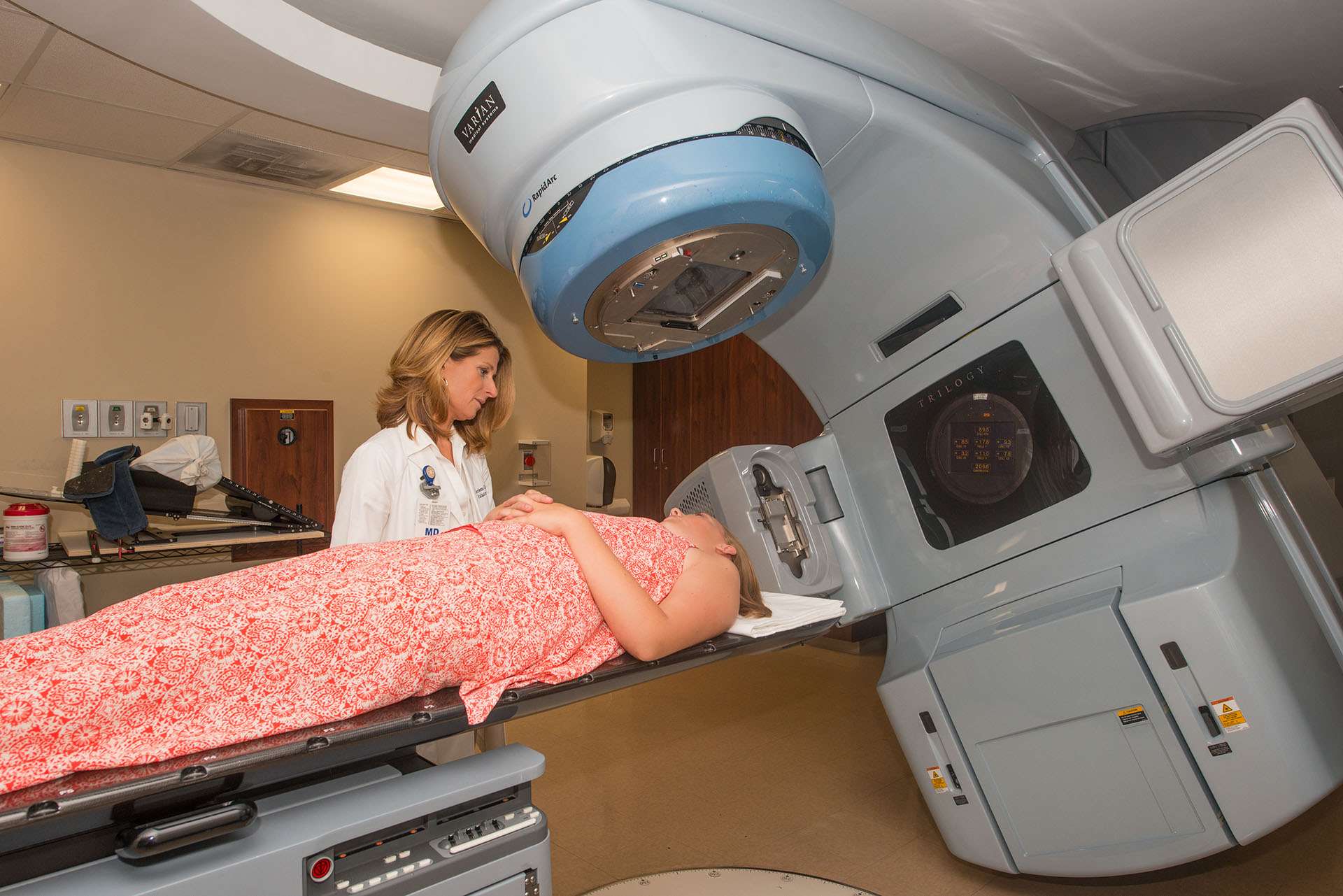
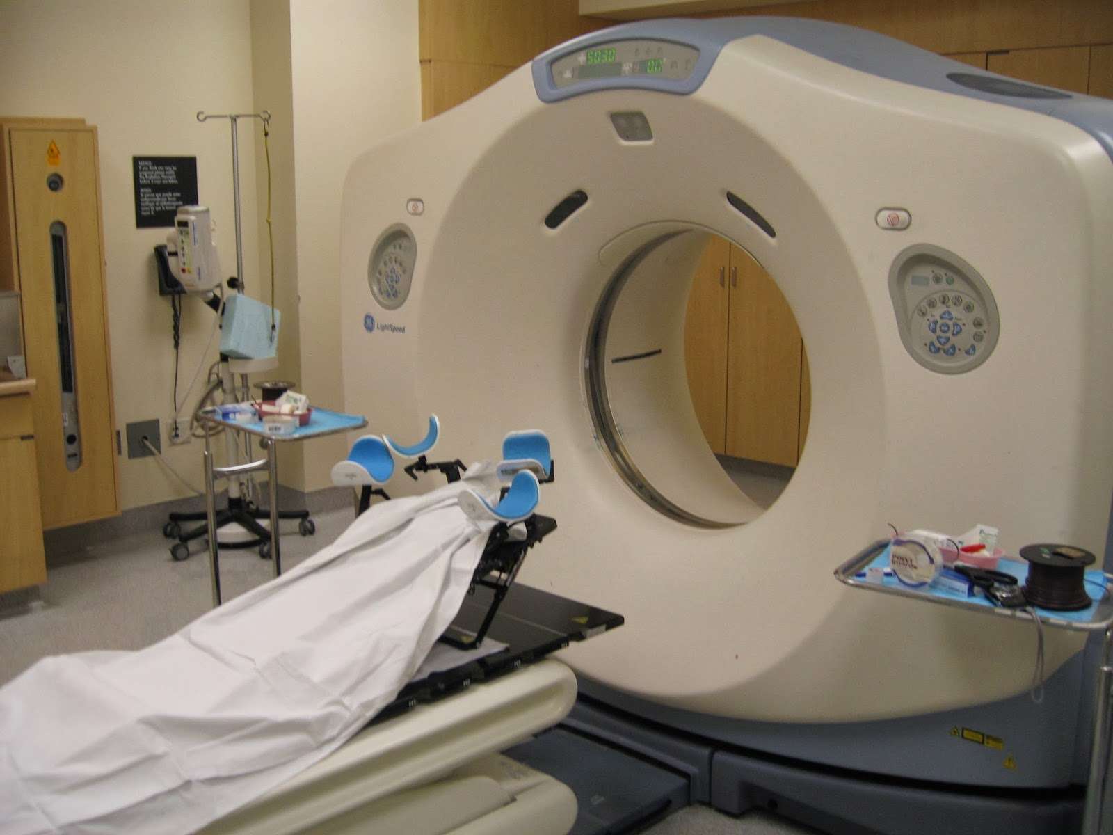
The SOMC Cancer Center uses a large bore CT scanner to produce detailed images of areas inside the body. These images are used by the radiation therapy planning team and your radiation oncologist to develop your customized treatment plan. The advanced imaging technology combines simulation and the respiratory motion to plan the radiation treatment with a high level of precision. There are many advantages to the patient in terms of use of this equipment:
- Accurately pinpoints tumor site or the site that the tumor was removed from while monitoring the motion of the area
- Enables the radiation oncologist to plan treatment in accordance with the patients breathing patterns
- Has a large opening to accurately scan patients of all sizes and accommodate positioning devices to assist the patient to hold still comfortably during the procedure
- Increased width of scanning space provides a more comfortable, less claustrophobic experience for patients
After Your Planning Session
You might have to wait a few days or up to 3 weeks before you start treatment. During this time your radiographers and doctors create a precise radiotherapy plan for you.
They make sure the treated area receives a high dose and nearby areas receive a low dose. This reduces the side effects you might get during and after treatment.
-
Scottish Intercollegiate Guidelines Network, September 2013
-
Postoperative radiotherapy for Breast Cancer: UK consensus statement
The Royal College of Radiologists, 2016
-
Early Breast Cancer: ESMO Clinical Practice Guidelines 2019F Cardoso and others
These Markings Are Done To Help Guide Treatment
Radiation can be an important part of treatment for breast cancer. During radiation treatment, high-energy beams are aimed at the breast tissue to kill cancer cells.
Before breast radiation is delivered, skin markings known as radiation tattoos, need to be placed on the skin. These marks help the radiation therapist aim the radiation precisely where it’s needed.
Radiation is typically given five days a week for about six weeks, and it’s critical the radiation is aimed at the same place in order to prevent cancer recurrence and to spare healthy tissue.
This article will review the process of tattoo placement and the types of breast tattoos available, as well as alternatives.
Don’t Miss: Does Soy Milk Cause Breast Cancer
Radiation Therapy: What To Expect
Many cancer patients will need radiation therapy as part of their treatment. Radiation can be used alone or as part of a treatment plan. When radiation is used in combination with other treatments, it can help to reduce the size of the tumor so that its easier to remove during surgery or to make it more sensitive to chemotherapy. Some patients also receive radiation after surgery or chemotherapy to help destroy any remaining cancer.
We spoke with radiation oncologist Bouthaina Dabaja, M.D., for insight on what patients can expect when receiving radiation treatment. Heres what she had to say.
How will I know if I need radiation therapy?
Your care team will work with you to determine your treatment plan, including whether you might need radiation therapy. If your care team determines that you need radiation therapy, then well decide the target, technique, dose and type of radiation to use.
What happens before radiation treatment begins?
What happens during the simulation process?
Depending on the cancer type, the consultation and simulation are usually scheduled on the same day. Before you start the simulation process, the therapists will explain the procedure and position you on the table. Simulation often takes 30-45 minutes.
Here are some other things to expect during the simulation:
Sexual And Reproductive Health

You can be sexually active during your radiation therapy unless your radiation oncologist gives you other instructions. You wont be radioactive or pass radiation to anyone else. If you or the person youre sexually active with can get pregnant, its important to use birth control during your radiation therapy.
You may have concerns about how cancer and your treatment can affect your sex life. Radiation therapy can affect your sexual health physically and emotionally. Talking with your radiation oncologist or nurse about your sexual health can be hard, but its an important conversation to have. They may not bring it up unless you share your questions and concerns. You may feel uncomfortable, but most people in cancer treatment have similar questions. We work hard to make sure everyone in our care feels welcome.
Sexual health programs
We also offer sexual health programs. These programs can help you manage the ways your cancer or cancer treatment affect your sexual health or fertility. Our specialists can help you address sexual health or fertility issues before, during, or after your treatment.
- For information about our Male Sexual & Reproductive Medicine Program or to make an appointment, call .
- For information about our Cancer and Fertility Program, talk with your healthcare provider.
Other resources
Recommended Reading: What Are Your Chances Of Getting Breast Cancer
During Your Radiation Therapy
On the day of your first radiation treatment, youll start putting triamcinolone 0.1% ointment on your skin in the treatment area. This is a prescription ointment that will help protect your skin. Youll use it every day, once in the morning and once in the evening. This includes the days you dont have treatment. Your radiation nurse will give you more information about it before your first treatment.
Your radiation oncologist may also recommend using Mepitel® Film to protect your skin in the treatment area. If they do, put it on your skin in the treatment area before your first treatment. Keep it on until the edges start to peel up.
Youll stay in one position for about 10 to 20 minutes during each of your radiation treatments, depending on your treatment plan. If you think youll be uncomfortable lying still, you can take acetaminophen or your usual pain medication 1 hour before your appointments.
Current Techniques In Three
Albert L. Wiley, Jr, MD, PhDOncology
The modern CT simulator is capable of interactive three-dimensional volumetric treatment planning this allows radiation oncology departments to operate without conventional x-ray simulators. Treatment planning is performed at the time of virtual simulation by contouring the organs or volumes of interest and determining the isocenter.
The modern CT simulator is capable of interactive three-dimensional volumetric treatment planning this allows radiation oncology departments to operate without conventional x-ray simulators. Treatment planning is performed at the time of virtual simulation by contouring the organs or volumes of interest and determining the isocenter. A digitally reconstructed radiograph provides a beam’s-eye-view display of the treatment field anatomy and contoured areas of interest. Conformal and noncoplanar teletherapy is facilitated for patients with prostate cancer, lung cancer, and brain tumors. Ongoing developments include 3D dose calculation, dose-volume histogram analysis, and tumor dose escalation.
You May Like: When To Get Tested For Breast Cancer
Side Effects Of Radiation Therapy To Your Breast Or Chest Wall
You may have side effects from radiation therapy. The type and how strong they are depends on many things. These include the dose of radiation, the number of treatments, and your overall health. The side effects may be worse if youre also getting chemotherapy.
You may start to notice side effects about 2 weeks after you start radiation therapy. They may get worse during your radiation therapy, but theyll slowly get better over 6 to 8 weeks after your last treatment. Some side effects may take longer to go away. Follow the guidelines in this section to help manage your side effects during and after your radiation therapy.
Tissue Density And Hu Management
The same curve to convert HU to mass density was used in PRIMO and Acuros based systems. The material assignment based on the CT number was set in PRIMO as similar as possible to the Acuros setting in Eclipse. Full compatibility of the two assignments is not viable, since Acuros assigns adjacent materials in a smooth way, allowing an overlapping HU range, where the previous and next materials are linearly combined from one to the other. The used materials are summarized in Table .
Table 1 HU and mass density ranges used in PRIMO and Acuros computations
One of the patients of this study was simulated with the two chemical compositions for adipose and muscle tissues, according to the PRIMO and Acuros defaults. With the PRIMO defaults, the dose to muscle and adipose tissues were estimated higher than using Acuros defaults by about 0.12% and 0.03, respectively. Those differences, although considered negligible, were excluded from the computation by changing the PRIMO tissue composition material defaults.
Recommended Reading: What Is The Main Cause Of Breast Cancer
Reslice Back With Virtual Seeds
One method of verifying the plan is by projecting the virtual implants back to their original data set. This innovative feature of our Vision system allows oncologists to view the implant position and spacing in 3D intuitively. In addition, Vision enables seed placement results to be viewed and validated.
Patient Doses With Monte Carlo Simulations
For each of the five cases, three different Monte Carlo simulations were computed in PRIMO, assigning different materials to the muscle and adipose HU ranges, while keeping the original density:
– AdiMus: as standard, muscle and adipose tissues were assigned to the muscle and adipose HU ranges, respectively
– Adi: the adipose tissue material was assigned to the HU including both adipose and muscle ranges
– Mus: the muscle tissue material was assigned to the HU including both adipose and muscle ranges.
Mean doses to CTV, CTV_lob and CTV_fat were computed for all the simulations.
The dose difference generated by the chemical composition of the specific tissue, lobular or fat, was estimated by the difference of the mean doses of the CTV_lob between Adi and AdiMus simulations, and of the difference of the mean doses of the CTV_fat between Mus and AdiMus simulations. Those values give the possible dose estimation error when a different material chemical composition is used for calculations, while the surrounding tissue dose is computed with the correct tissue assignment. Calculations were based on the mean dose of the whole structure. Uncertainties were reported at two standard deviations for all the voxels in each specific structure.
Read Also: Can You Die From Stage 4 Breast Cancer
When To Call Your Radiation Oncologist Or Nurse
- You have a fever of 100.4 °F or higher.
- You have chills.
- Your skin is painful, peeling, blistering, moist, or weepy.
- You have discomfort in the treatment area.
- Your breast, underarm , or arm is getting more swollen.
- You have any new or unusual symptoms.
Many people find counseling helpful. We provide counseling for individuals, couples, families, and groups. We can also prescribe medications to help if you feel anxious or depressed. To make an appointment, ask your healthcare provider for a referral or call the number above.
Integrative Medicine Servicewww.mskcc.org/integrativemedicineOur Integrative Medicine Service offers many services to complement traditional medical care, including music therapy, mind/body therapies, dance and movement therapy, yoga, and touch therapy. To schedule an appointment for these services, call .
You can also schedule a consultation with a healthcare provider in the Integrative Medicine Service. They will work with you to come up with a plan for creating a healthy lifestyle and managing side effects. To make an appointment, call .
Nutrition ServicesOur Nutrition Service offers nutritional counseling with one of our clinical dietitian nutritionists. Your clinical dietitian nutritionist will talk with you about your eating habits. They can also give advice on what to eat during and after treatment. To make an appointment, ask a member of your care team for a referral or call the number above.
For more information, call the number above.
Clinical Results Of Conformal Radiation Therapy

Prostate Cancer
Investigators have utilized conformal radiation therapy for the treatment of localized prostate cancer. Soffen, Hanks, and colleagues reported a reduction in acute morbidity with conformal therapy, as compared with nonconformal techniques . Hanks et al further noted an average 14% reduction in rectal and bladder dose exposure with conformal therapy. Their experience using conformal therapy in 108 patients with localized prostate carcinoma revealed only 1% with Radiation Therapy Oncology Group grade 3 or 4 complications over a median follow-up of 16 months . A recent report on this experience showed a 30% reduction in grade 2 urinary complications in the group receiving conformal therapy, as compared with a conventionally treated control group. Dose escalation to 75 to 79 Gy was achieved in 20 patients .
Leibel and coworkers described dose escalation using conformal therapy for localized prostate carcinoma in 123 patients. Doses ranged from 64.8 to 75.6 Gy. Only one patient experienced RTOG grade 4 toxicity, 67% of patients had normalized serum prostate-specific antigen levels within 14 months of treatment , and the early disease-free rate was 89% .
Lung Cancer
Other Carcinomas
An NCI multi-institutional dose escalation study is currently evaluating the patient tolerance to higher doses of irradiation given with conformal techniques.
Read Also: How Can Guys Get Breast Cancer
D Ct Simulation Procedure
CT Data Acquisition
Computed tomographic scanning is performed with a 70-cm ring aperture and flat couch, which is integrated into a computer-based virtual simulator capable of 3D volumetric reconstruction and multiplanar reconstruction for treatment planning. A laser isocenter projection system is mounted in the room the system combines two fixed and one movable sagittal laser with a variance of only 2 mm over a 6-month period. Digitally reconstructed radiographs with superimposed target volume information are generated by the computer and transferred to a commercially available CT film processor. Analysis of DRRs reveals acceptable bone detail and anatomic agreement with port films taken on the treatment unit.
The best quality DRRs are obtained with a nonspiral CT slice thickness of 2 mm and table increment of 2 mm. A slice thickness of 5 mm and table increment of 3 mm also yield DRRs of excellent quality. For larger fields up to 40 cm in length, a 5-mm slice with 5-mm table increment is used most commonly, and results in lesser detail but clinically acceptable positioning accuracy and results. For patients who cannot stay in position for more than 15 to 20 minutes, a 10-mm slice and 10-mm table increment provide acceptable quality for treatment of bone metastases and other simple treatment set-ups. Plastic mask immobilization facilitates brain, head, and neck positioning and stability.
Treatment Planning
Digitally Reconstructed Radiograph
3D Dose Calculations
Proton Beam Radiation Therapy
Proton beam radiation uses beams of protons instead of X-rays. A proton is a particle with a positive electric charge that is in the nuclei of all atoms.
X-rays release energy both before and after they hit their target. But protons release their energy only after traveling a certain distance. So doctors think protons may be able to deliver more radiation directly to the treatment area while possibly doing less damage to nearby healthy tissue. But this is still being studied.
Right now, proton beam radiation is only being used in clinical trials to treat breast cancer. The machines needed to deliver protons are very expensive and are not widely available.
If youre interested in being treated with proton beam therapy, talk to your doctor to find a clinical trial that would be a good fit for your unique situation.
You May Like: Can You Get Breast Cancer From Sleeping In Your Bra
Current Fractionated Radiotherapy Technique
The planning process for fractionated radiotherapy begins with a CT-based simulation. During the CT simulation, the patient is placed supine on the CT simulator couch and a thermoplastic mask or other, highly accurate, immobilization system is fitted to the patient. A CT scan with slice thickness varying from 0.5 mm to 2.5 mm is then performed with the patient immobilized and wearing the thermoplastic mask in the treatment planning position.
The use of MRI in the treatment planning process is, for the most part, a requirement in order to effectively delineate the tumor and define normal structures. A high-resolution axial MRI with and without gadolinium contrast is generally performed either a few days before or after a CT simulation and then registered to the CT simulation using specialized software.
The PTV is then treated to a prescription dose of 5054 Gray in standard fractionation . Given the generally young age of patients and proximity to critical structures, it is recommended that a high conformality radiation delivery technique be employed, including, but not limited to, IMRT, tomotherapy, or volumetric modulated arc therapy . In rare instances in which such modern techniques are not available, three-dimensional conformal radiation therapy could be used but would be considered by many radiation oncologists to be sub-optimal. Margins, of course, would need to be adjusted for 3D-CRT.
Sonja Dieterich PhD, DABR, … Jing Zeng MD, DABR, in, 2016
Definitions For Model Classification
During the data analysis, we compiled several keywords describing simulation approaches and models used by authors to describe their models. Several entries were combined to simplify the classification and some conceptually similar simulation approaches were merged. The full classification unification table can be found in Additional file 2: Appendix D . We identified four main simulation approaches that applied to 87% of considered studies.
A cohort-level model refers to a Markov chain model used to calculate the transitions from one population group to another.
An individual-level model is a Markov chain model that calculates transitions between health states for an individual. The most popular name for such models is microsimulation, but not all authors have consistently used this term .
Regression models cover all types of regression : e.g., linear models, generalized linear and non-linear models, multivariate models, and mixed-effects models. This group includes all cohort and individual level regressions.
Differential equations models can also be linear, non-linear, or partial, etc. This group includes all individual and cohort level DEs.
We classified studies as “applied” if the model was used to obtain the study’s main result, and “developed” if the paper’s main outcome was the model itself, and the model is described in sufficient detail to be reproduced. Studies that could not be classified into one of the two above mentioned categories were labeled as “Other.”
Recommended Reading: Does Breast Cancer Cause Bruising