Phyllodes Tumor Of The Breast
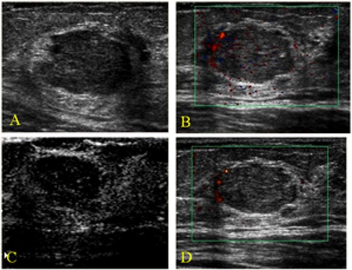
This 48 year of female patient had a large lump in the medial aspect of the left breast. The ultrasound images of the breast show a large inhomogenous mass of 5.6 x 3.4 cms. with multiplelobulations and cystic spaces also present. The appearance of the tumor was leaf like in its internal architecture. There is also posterior acoustic enhancement.
The images above show minimal internal vascularity on color Doppler ultrasound and 3-D ultrasound , show the internal lobulation with typical leaf like pattern on sonography. The ageof the patient, large size of tumor and typical ultrasound features are highly suggestive of this being a Phyllodes tumor of the left breast. Ultrasound images of Phyllodes tumor are courtesy of RaviKadasne, MD, UAE.
Characteristics Of Malignant Lesions
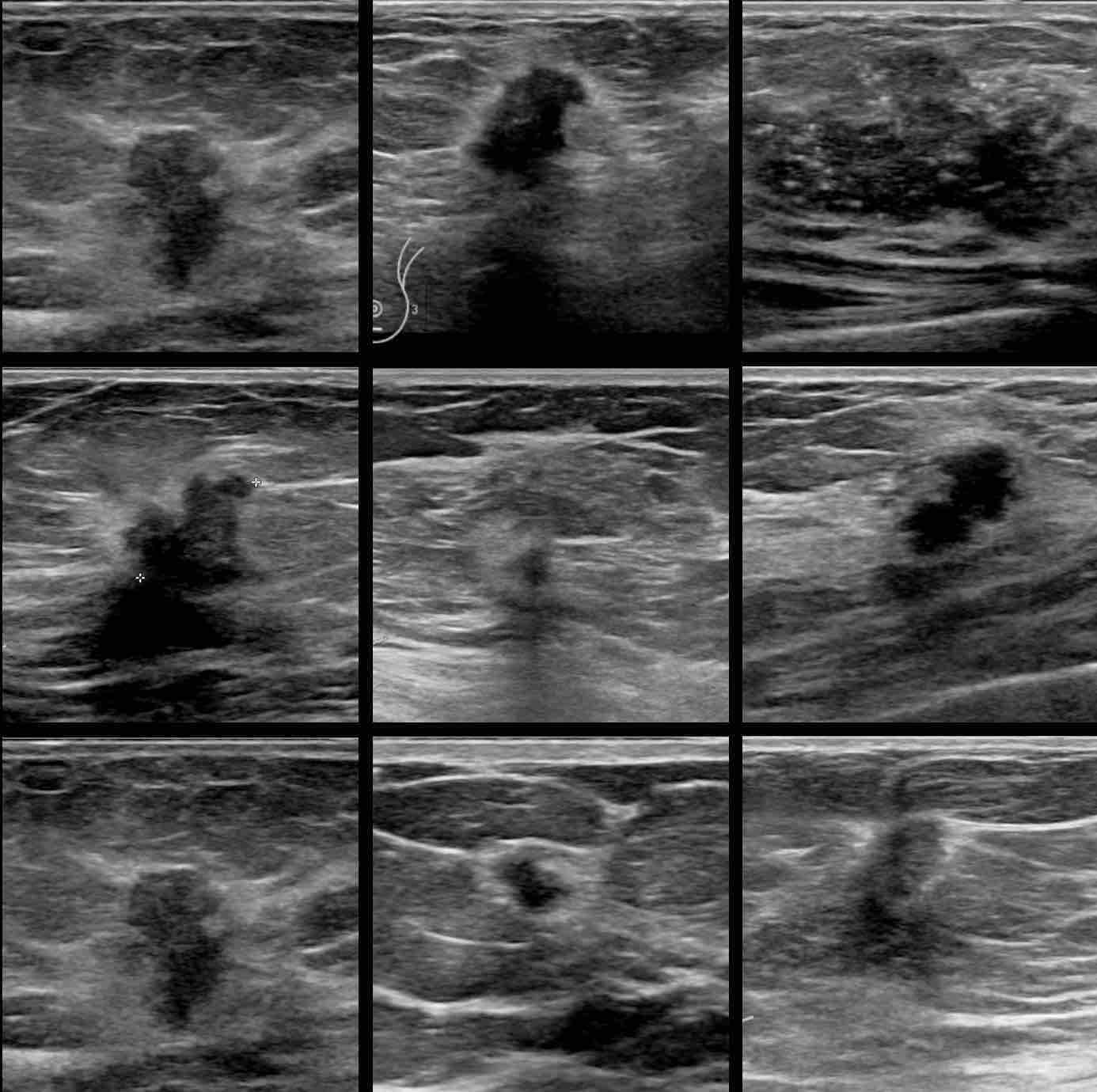
Malignant lesions are commonly hypoechoic lesions with ill-defined borders. Typically, a malignant lesion presents as a hypoechoic nodular lesion, which is taller than broader and has spiculated margins, posterior acoustic shadowing and microcalcifications . Three-dimensional scanners with the capability of reproducing high-resolution images in the coronal plane provide additional important information. The spiky extensions along the tissue planes can be well seen in coronal images .B]. It was initially believed that color Doppler scanning would add to the specificity of USG examination, but this has not proven to be very efficacious however, in certain situations it does help resolve the issue, particularly when there is significant vascularity present within highly cellular types of malignancies .
Malignant lesions. Transverse scan shows a typical malignant nodule that is taller than wide, with hypoechoic echotexture. Arrowheads indicate irregular spiculated margins. Some of the nodules may reveal a branching pattern . Sagittal view shows a nodule with multilobulated margins the presence of more than 34 lobulations is suspicious for malignancy. Sagittal and transverse scans show duct extension . M indicates the primary site. Duct extension appears smooth in outline in cross-section . Transverse scan shows a typical malignant lesion with irregular spiky margins, microcalcifications and a branching pattern. This lesion is classifiable as US-BIRADS category 4
Screening And Detection Rates Using Ultrasound
The discovery rate of malignant cancer using breast cancer screening methods is actually very low.
So, for example, the rate of detecting breast cancer on a mammogram is about 5 cancers in every thousand women screened.
Ultrasound alone for breast cancer screening detects slightly fewer malignancies. This tends to suggest that mammography is slightly more reliable in the detection of breast cancer.
Unfortunately, I have to stress here that the combination of mammography, ultrasound and even MRI can not completely exclude the possibility of breast cancer.
Indeed, up to 3% of women with suspicious lesions but negative mammograms and ultrasounds may still have breast cancer.
Don’t Miss: Breast Cancer Stage 1 Chemotherapy
How Does An Ultrasound Help To Diagnose A Breast Lump
When a suspicious site is detected in your breast through a breast self-exam or on a screening mammogram, your doctor may request an ultrasound of the breast tissue. A breast ultrasound is a scan that uses penetrating sound waves that do not affect or damage the tissue and cannot be heard by humans. The breast tissue deflects these waves causing echoes, which a computer uses to paint a picture of whats going on inside the breast tissue . A mass filled with liquid shows up differently than a solid mass.
How Is A Breast Ultrasound Done

Most often, ultrasound is done using a handheld, wand-like instrument called atransducer. First a gel is is put on the skin and/or the transducer, and then the transducer is moved around over the skin. It sends out sound waves and picks up the echoes as they bounce off body tissues deeper under the skin. These echoes are made into a picture on a computer screen. You might feel some pressure as the transducer is moved around on your skin, but it should not be painful.
Automated breast ultrasound is an option at some imaging centers. This technique uses a much larger transducer to take hundreds of images that cover nearly the entire breast. ABUS might sometimes be done as an added screening exam for women who have dense breasts. It might also be used in women who have abnormal findings on other imaging tests or who have breast symptoms. When ABUS is done, a second handheld ultrasound is often needed to get more pictures of any suspicious areas.
Also Check: Can You Recover From Stage 4 Breast Cancer
What Are The Signs And Symptoms Of Breast Cancer
Knowing the signs and symptoms of breast cancer may help save your life. When the disease is discovered early, there are more treatment options and a better chance for a cure.
Most painful breast lumps are not cancerous. Any discrete breast lump whether painful or not should be evaluated because breast cancer often presents as a lump or thickening.
How Does The Procedure Work
Ultrasound imaging uses the same principles as the sonar that bats, ships, and fishermen use. When a sound wave strikes an object, it bounces back or echoes. By measuring these echo waves, it is possible to determine how far away the object is as well as its size, shape, and consistency. This includes whether the object is solid or filled with fluid.
Doctors use ultrasound to detect changes in the appearance of organs, tissues, and vessels and to detect abnormal masses, such as tumors.
In an ultrasound exam, a transducer both sends the sound waves and records the echoing waves. When the transducer is pressed against the skin, it sends small pulses of inaudible, high-frequency sound waves into the body. As the sound waves bounce off internal organs, fluids and tissues, the sensitive receiver in the transducer records tiny changes in the sound’s pitch and direction. A computer instantly measures these signature waves and displays them as real-time pictures on a monitor. The technologist typically captures one or more frames of the moving pictures as still images. They may also save short video loops of the images.
Doppler ultrasound, a special ultrasound technique, measures the direction and speed of blood cells as they move through vessels. The movement of blood cells causes a change in pitch of the reflected sound waves . A computer collects and processes the sounds and creates graphs or color pictures that represent the flow of blood through the blood vessels.
Also Check: When Can Breast Cancer Start
Is Breast Ultrasound Used In Breast Cancer Screening
Ultrasound imaging is not typically used in the detection of breast cancer by itself, as it may not pick up some of the earlier signs of the disease. For example, tiny deposits of calcium are easily visible in mammograms but usually do not show up on ultrasound images. In many cases, calcium deposits are benign , but they can also be an early marker for breast cancer.
Instead, ultrasound is used alongside other breast cancer screening tests. As one MyBCTeam member shared, I never felt a lump or had any tenderness in my breasts. Ultrasounds picked it up when it first looked suspicious, and then they followed it for a few years. When it continued to grow and spread, they did the biopsies.
If you detect a change in your breast, such as a new lump, during a self-exam or mammography, a breast ultrasound can provide your health care team with more information about the change. The test can help determine whether a breast lump is solid or filled with fluid . It is important to note that ultrasound on its own cannot detect whether a solid mass in the breast is cancerous or benign.
Will My Doctor Call Me With Ultrasound Results
You may be told the results of your scan soon after its been carried out, but in most cases the images will need to be analysed and a report will be sent to the doctor who referred you for the scan. Theyll discuss the results with you a few days later or at your next appointment, if ones been arranged.
Read Also: What Does Breast Cancer Rash Look Like
How Is Breast Ultrasound Used
A breast ultrasound may be used in several ways. In many cases, it helps reveal breast abnormalities that mammography cannot easily detect. Although older womens breasts tend to have a fattier composition , younger womens breasts are often denser. Dense breast tissues may resemble cancerous tumors on a mammogram. Ultrasound can help detect breast lumps through this dense tissue more clearly than a mammogram.
What Will I Experience During And After The Procedure
Most ultrasound exams are painless, fast, and easily tolerated.
Breast ultrasound is usually completed within 30 minutes.
If the doctor performs a Doppler ultrasound exam, you may hear pulse-like sounds that change in pitch as they monitor and measure the blood flow.
You may need to change positions during the exam.
When the exam is complete, the technologist may ask you to dress and wait while they review the ultrasound images.
After an ultrasound exam, you should be able to resume your normal activities immediately.
Read Also: Can You Get Breast Cancer At 24
How Are Breast Ultrasound Results Reported
Doctors use the same standard system to describe results of mammograms, breast ultrasound, and breast MRI. This system sorts the results into categories numbered 0 through 6.
For more details on the BI-RADS categories, see Understanding Your Mammogram Report. While the categories are the same for each of these imaging tests, the recommended next steps after these tests might be different.
What Does Ultrasound Show That Mammogram Doesn T

Mammograms: If a screening mammogram identifies a suspicious area in the breast, you will likely need a diagnostic mammogram. A diagnostic mammogram takes more pictures than a routine screening mammogram and focuses on the affected area. Ultrasounds: A breast ultrasound cannot spot microcalcifications in the breast.
Recommended Reading: What Is Oncotype Dx Test For Breast Cancer
What Does Breast Cancer Look Like In An Ultrasound
Fluid-filled cysts usually appear as solid black circles or ovals, while compared to cysts, breast cancers usually appear as slightly lighter irregular masses. It is important, however, not to try to interpret breast cancer ultrasound images on your own. Your healthcare professional is trained to know what to look for and will talk you through any next steps.
Breast cancer shows up as a darker patch on the otherwise lighter grey or white tissue around it. This is because the sound waves will bounce off a solid tumour. Surrounding tissue will be interpreted by the computer as a lighter-coloured image with the tumour mass appearing darker than surrounding healthy tissue such as air and bone.
An ultrasound can detect potential breast cancers, however, it cannot determine whether a solid lump is cancerous or not only whether a lump is solid , filled with fluid or both. An ultrasound may also fail to detect small lumps and cannot pick up small deposits of calcium that can indicate breast cancer. If a solid, potentially cancerous tumour appears in your breast cancer ultrasound, your doctor will require further testing for a complete diagnosis.
For breast cancer, an ultrasound is highly accurate, with research suggesting it can correctly detect around 80% of breast cancers. It is useful in the detection and diagnosis of breast cancer, but as it cannot indicate whether a solid mass in the breast is cancerous or benign, it is usually used alongside other tests.
S Of Breast Cancer Lumps: Early Breast Cancer Signs To Look For
For half the population, Breast Cancer Awareness Month in October means being bombarded with pictures of breast cancer lumps, and messages encouraging them to “be familiar” with their breasts and know the warning signs of breast cancer. However, what’s normal for one pair of breasts may not be for another, so it can be tricky to determine what’s just typical variation and what’s a warning sign.
TODAY spoke to several experts who explained what to be aware of and when you should see a doctor.
Recommended Reading: What Stage Is Metastatic Breast Cancer
Can I Get A Breast Ultrasound Instead Of Mammogram
A breast ultrasound isnt typically a screening tool for breast cancer. Instead, a physician might order an ultrasound, also called a sonogram, of the breasts if a screening mammogram produces unusual results. A physician might also use a breast ultrasound as a visual guide while performing a biopsy of the breasts.
Which Is Better Breast Mri Or Ultrasound
Screening via MRI is a good idea, especially for women at high risk who are getting surgery on one breast, says Hryniuk. Even for large tumours, MRIs have been shown to be more accurate than physical exams, mammography or ultrasounds in following the results of chemotherapy to shrink large breast tumours, Hryniuk says.
Recommended Reading: When You Have Breast Cancer Does It Hurt
Normal Breast Parenchymal Patterns
In the young non-lactating breast, the parenchyma is primarily composed of fibroglandular tissue, with little or no subcutaneous fat. With increasing age and parity, more and more fat gets deposited in both the subcutaneous and retromammary layers .
Normal breast. Mid transverse scan of a normal breast. The fibroglandular parenchyma is echogenic and is surrounded by hypoechoic fat
Color Doppler Sonography: Characterizing Breast Lesions
Atif Hashmi, Susan Ackerman and Abid Irshad
Department of Radiology, Medical University of South Carolina, PO Box 250322, 169 Ashley Avenue, Charleston, SC 29425, USA
- *Corresponding Author:
- Department of Radiology, Medical University of South Carolina PO Box 250322, 169 Ashley Avenue CharlestonSC 29425, USA Tel: +1 843 792 1957 Fax:+1 843 792 9503 E-mail:
You May Like: Olivia Newton John Breast Cancer
Benign Indeterminate And Malignant Nodules
In a landmark study, Stavros et al established US criteria for characterizing solid breast masses. This work was facilitated by evolving technical improvements in US equipment that provided better resolution and images. They demonstrated that US may be used to accurately classify some solid lesions as benign, allowing follow-up with imaging rather than biopsy. They used high-resolution transducers, which were state-of-the-art at that time, and performed examinations in both radial and antiradial planes. The investigators also focused on the evaluation of suspected areas in the transverse and longitudinal planes.
Stavros et al proposed a US scheme for prospectively classifying breast nodules into 1 of 3 categories :
To be classified as benign, a nodule had to have no malignant characteristics. In addition, 1 of the following 3 combinations of benign characteristics had to be demonstrated:
-
Intense uniform hyperechogenicity
-
Ellipsoid or wider-than-tall orientation, along with a thin, echogenic capsule
-
2 or 3 gentle lobulations and a thin, echogenic capsule
A nodule was classified as indeterminate by default if it had no malignant characteristics and none of the 3 benign characteristic combinations listed above.
To be classified as malignant, a mass needed to have any of the following characteristics:
-
Spiculated contour
-
Microlobulation
Why Do Doctors Order Ultrasounds

For example, they may order an ultrasound to check a lump that a person can feel during a physical examination but that a mammogram does not detect. The ultrasound image can also help a doctor distinguish between a solid mass and a fluid-filled sac, which healthcare professionals refer to as a cyst.
You May Like: How Many Stages Are There To Breast Cancer
The Reports Of Your Radiology Exams Usually Contain Three Sections:
- Exam description and history the type of exam, day it was performed, the reason it was performed and any important patient information
- Findings a detailed description of the important findings on the exam including size, shape, location and changes
- Impression a summary of the findings, what they mean and what to do about them Radiologists use standard terms in reports to describe the appearance of important findings.
Some examples of those terms include mass, architectural distortion and calcifications. The radiologist will also describe the size, shape and location of important findings. The size and location can be critical to making decisions about the kind of operation and other treatments you might have.
Radiologists will use a clock face or quadrant to describe the location. There is a separate clock for each breast and they are oriented as if the doctor is looking at you during an examination. In the diagram below, the nipple is in the center of the clock for both breasts. The outer left breast is at 3 oclock and the outer right breast is at 9 oclock. In the left breast the upper outer quadrant is between 12 and 3 oclock.
The radiologist will also describe the size and location of a finding by indicating the distance from the nipple in centimeters. Centimeters are smaller than an inch. There are 2.54 centimeters in an inch.
For example:
How Is The Procedure Performed
You will lie on your back or on your side on the exam table. The sonographer may ask you to raise your arm above your head.
The radiologist or sonographer will position you on the exam table. They will apply a water-based gel to the area of the body under examination. The gel will help the transducer make secure contact with the body. It also eliminates air pockets between the transducer and the skin that can block the sound waves from passing into your body. The sonographer places the transducer on the body and moves it back and forth over the area of interest until it captures the desired images.
There is usually no discomfort from pressure as they press the transducer against the area being examined. However, if the area is tender, you may feel pressure or minor pain from the transducer.
Doctors perform Doppler sonography with the same transducer.
Once the imaging is complete, the technologist will wipe off the clear ultrasound gel from your skin. Any portions that remain will dry quickly. The ultrasound gel does not usually stain or discolor clothing.
You May Like: Do Obgyn Check For Breast Cancer