What If My Report Mentions Micrometastases In A Lymph Node
This means that there are cancer cells in the lymph nodes that are bigger than isolated tumor cells but smaller than regular cancer deposits. If micrometastases are present, the N category is described as pN1mi. This can affect the stage of your cancer, so it might change what treatments you may need. Talk to your doctor about what this finding may mean to you.
Read Also: Stage Four Breast Cancer Prognosis
How Do You Count Mitotic Counts
Mitotic count is determined most frequently in veterinary pathology, usually at 400 magnification, but the area counted is seldom defined, and the term often reported is mitotic index. Mitotic index is defined as the number of cells undergoing mitosis divided by the number of cells not undergoing mitoses.
Additional Grading Criteria: A Composite Total Of Tubular Nuclear And Mitotic Index Assesments
As a grade of low, intermediate or high is obtained through a composite sum by assigning a score based on the nuclear assessment, a mitotic index assessment, and a tubular assessment.
The nuclear assessment is based on the nuclear size within the invasive cells. They are described from small to medium to large in size, as well as by their uniformity in size and shape.
The tubular assessment refers to an approximate, quantitative account of the amount of cell groupings which remain in their normal tubular shape. The smaller the percentage of tubular structures in comparison to other shapes, the higher the score. Other structures to appear may include solid trabecula, vacuolated single cells, alveolar nests, and solid sheets of cells.
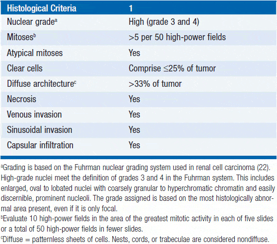
The mitotic index refers to evident patterns of cell division.Mitosis is a process by which a cell separates into two genetically identical daughter cells. . So, the mitotic index is assessment of the abundance of these pairs of daughter cells, measured in the count per square millimeter. Mitoses are only counted in the invasive area of the lesion .
| Histologic Grade | |
| 11-19 = 2 | > 20 = 3. |
Recommended Reading: Estrogen Positive Breast Cancer Recurrence
What Is The Meaning Of Mitotic
: of, relating to, involving, or occurring by cellular mitosis mitotic cell division mitotic recombination Microtubules move material through the cell and, in particular, form an important component of the mitotic spindle, which is a structure that separates the duplicated sets of chromosomes in the course of cell
What Does It Mean If My Doctor Asks For A Special Molecular Test To Be Performed On My Specimen

Molecular tests such as Oncotype DX® and MammaPrint® may help predict the prognosis of certain breast cancers, but not all cases need these tests. If one of these tests is done, the results should be discussed with your treating doctor. The results will not affect your diagnosis, but they might affect your treatment.
Recommended Reading: Is It Possible To Have Breast Cancer At 13
The Pathology Report Is A Collection Of Information That Describes A Patients Breast Cancer
- How aggressive is the breast cancer?
- Have any cancer cells left the original tumor and traveled elsewhere, such as the underarm lymph nodes? Are they likely to travel?
- What determines if my cancer will respond to treatment?
Pathologists are doctors responsible for looking at your tissue sample under the microscope. This allows them to assess the cells for abnormalities that could lead to the diagnosis of breast cancer. They prepare a report about their findings. The report has information about the size, shape and appearance of the cancer as it looks to the naked eye. Pathology reports play an integral role in the diagnosis of breast cancer and staging.
Using Conventional Microscopes With Different Sized Hpf
It is a common practice to count mitotic figures in 10 HPF at ×40 magnification in several fields of pathology, including breast but in certain tumours where MC can be highly variable, more HPF are required to account for this variability. For instance, in adrenal gland and gastrointestinal stromal tumours, mitotic figures are counted in 50 HPF or in 5mm2 areas.
Although a gradual firm move has been made towards standardising the area of measurement, HPF is still widely used without score adjustments throughout different fields of pathology apart from BC and few other tumours.
Adjustment of mitotic scores based on the field diameter of the microscopes only provides standardisation of counting and may improve the concordance of scores when using the same microscopes, but does not address the clinical impact of the MC identified or the concordance when using different microscopes with variable field diameters. Theoretically and even in practice, MC in 10 HPF, which is usually equal to 22.5mm2 in most modern used microscopes are not reliable.
Even the original Nottingham grade authors have mentioned the need to count more than 10 HPF, but it has not been sufficiently highlighted, and only by increasing the sample size, this problem can be addressed.
Also Check: What Does Metastatic Breast Cancer Mean
How Do Cancer Cells Travel
Cancer cells use the lymph system as a first step to traveling to other areas of the body. During a breast cancer surgery, lymph nodes are removed and checked for the presence of cancer cells. This will be reported as the number of lymph nodes that contained cancer cells and how many were examined. For example, the report might state ten benign lymph nodes or tumor seen in ten of twelve lymph nodes .
What Is Histologic Grade Or Nottingham Grade Or Elston Grade
These grades are similar to what is described in the question above about differentiation. Numbers are assigned to different features seen under the microscope and then added up to assign the grade.
- If the numbers add up to 3-5, the cancer is grade 1 .
- If they add up to 6 or 7, it means the cancer is grade 2 .
- If they add up to 8 or 9, it means the cancer is grade 3 .
Also Check: Are There Prenatal Tests For Breast Cancer
What If My Report Mentions Her2/neu Or Her2
Some breast cancers have too much of a growth-promoting protein called HER2/neu . The HER2/neu gene instructs the cells to make this protein. Tumors with increased levels of HER2/neu are referred to as HER2-positive.
The cells in HER2-positive breast cancers have too many copies of the HER2/neu gene, resulting in greater than normal amounts of the HER2 protein. These cancers tend to grow and spread more quickly than other breast cancers.
All newly diagnosed breast cancers should be tested for HER2, because women with HER2-positive cancers are much more likely to benefit from treatment with drugs that target the HER2 protein, such as trastuzumab , lapatinib , pertuzumab , and T-DM1 .
Testing of the biopsy or surgery sample is usually done in 1 of 2 ways:
- Immunohistochemistry : In this test, special antibodies that will stick to the HER2 protein are applied to the sample, which cause cells to change color if many copies are present. This color change can be seen under a microscope. The test results are reported as 0, 1+, 2+, or 3+.
- Fluorescent in situ hybridization : This test uses fluorescent pieces of DNA that specifically stick to copies of the HER2/neu gene in cells, which can then be counted under a special microscope.
Many breast cancer specialists think that the FISH test is more accurate than IHC. However, it is more expensive and takes longer to get the results. Often the IHC test is used first:
Pathological And Molecular Characterization Of Triple Negative Breast Cancer
As a distinct molecular entity, TNBCs appear to be quite heterogeneous at a histopathological level. They frequently show features of ductal invasive carcinomas although metaplastic, medullary and apocrine features are also found . Moreover, TNBCs may present themselves as adenoid cystic lesions, histiocytoid carcinomas and even as invasive lobular carcinomas . A relatively large number of breast cancers that do not exhibit a basal phenotype appeared to have a triple-negative profile . Therefore, from a morphological and molecular point of view, TNBC may somehow be classified into four main categories that include the normal-like and the apocrine subtypes . It appears that some histopathological features of breast cancer such as pleomorphic lobular carcinoma and mixed ductal-lobular carcinoma exhibit a triple-negative molecular profile . Also, most invasive carcinomas that develop from microglandular adenosis areas are in fact triple-negative tumors . Recent studies have shown that metaplastic carcinomas are usually TNBCs . Metaplastic carcinomas are known to be rare, aggressive diseases of the breast that are usually diagnosed at grade 3 and, similar to TNBCs, have no specific therapeutic guidelines .
Don’t Miss: Symptoms Of Inflamatory Breast Cancer
What Is The Pathologic Stage Of Cancer
The pathologic stage of a cancer takes into consideration the characteristics of the tumor and the presence of any lymph nodes metastases or distant organ metastases . These features are assigned individual scores called the pathologic T stage , N stage and M stage are combined to form a final overall pathology stage . The pathologic stage is determined by the findings at the time of surgery and is different from the clinical stage, which is the stage estimated based upon the findings on clinical exam and radiology.
What Is Tumor Grade In 2021

Updated on March 29, 2021. Tumor grade is one of many items that will appear on your pathology report if you have breast cancer. It is a description of what the cell looks like under the microscope, the characteristics of which can tell a doctor how likely it is to grow and spread. Knowing the tumor grade can play a key role in helping your doctor
Recommended Reading: What Does Breast Cancer Do To The Body
Of Navigating Wsis When Counting Mitoses
To select the most reliable approach for counting mitotic figures on WSI, we compared three methods including multiple display screens at ×40 magnification screen fields ) equivalent to of 3mm2 area . counting within a pre-annotated as a single area. counting within a pre-annotated as multiple separate rectangles (non-adjacent small areas in different hotspots to avoid areas with low cellularity or artifacts that collectively equivalent to 3mm2. Measurement of accuracy is challenging as no ground truth is available to compare each method against. We have checked the accuracy by annotating and re-assessing some of the figures that were detected by one method and missed by the others, and they were agreed by the two observers to be true mitotic figures high concordance was used as evidence of acceptance. One of the main aims for the choice of the method is the reproducibility of the technique, as well as the consistency and concordance of scoring. Other variables include time, pathologists preferences, matching with the existing guidelines and current practice.
What If My Report Mentions Lymph Nodes
If breast cancer spreads, it often goes first to the nearby lymph nodes under the arm . If any of your underarm lymph nodes were enlarged , they may be biopsied at the same time as your breast tumor. One way to do this is by using a needle to get a sample of cells from the lymph node. The cells will be checked to see if they contain cancer and if so, whether the cancer is ductal or lobular carcinoma.
In surgery meant to treat breast cancer, lymph nodes under the arm may be removed. These lymph nodes will be examined under the microscope to see if they contain cancer cells. The results might be reported as the number of lymph nodes removed and how many of them contained cancer .
Lymph node spread affects staging and prognosis . Your doctor can talk to you about what these results mean to you.
You May Like: Breast Cancer In 20 Year Olds
Mai Assessment On Wsi
Glass slides were scanned using a Leica Scanner SCN400 at 40Ã. The standard image viewer for Leica Scanner Digital Image Hub was used for annotating and exploring WSI. WSI were displayed on high resolution 30 Barco Pathology Displays having a resolution of 6 MP. Examining WSI on 40Ã, in an area of 2 mm2, about 7 screen fields fitted into the same 2 mm2 area annotated before on the glass slides. Each observer was asked to annotate all the mitotic figures that he could detect within this area. Afterwards mitoses annotations were counted for each observer separately. Figure 1 is a snapshot from a WSI of an invasive breast cancer showing the selected areas for counting mitosis digitally and microscopically in addition to the digitally annotated mitotic figures within a 2 mm2 area.
What Hormones Make My Cancer Grow
Hormone Receptor StatusIf your cancer cells have a high proportion of estrogen or progesterone receptors, the report will say you are ER positive or PR positive. If your cells have a lower number of receptors, your report will say you are ER or PR negative. Another way to think of this is a car and driver example. The hormone buckles itself into the car seat to drive the tumor to make it grow. This is one of the most important pieces of information on the pathology report. Being ER/PR positive means you might benefit from hormonal therapy. Hormone therapy is actually therapy with an oral drug, usually Tamoxifen or aromatase inhibiters, which blocks hormone receptors in the cancer cell.
Recommended Reading: Final Stages Of Breast Cancer
Don’t Miss: Does Breast Cancer Make You Cough
What Is Mitotic Rate And Tubule Formation
Mitotic Rate: Describes how quickly the cancer cells are multiplying or dividing using a 1 to 3 scale: 1 being the slowest, 3 the quickest. Tubule formation: This score represents the percent of cancer cells that are formed into tubules. A score of 1 means more than 75% of cells are in tubule formation.
Mortality Rates Versus Number Of Breast Cancer Deaths
Sometimes its useful to have an estimate of the number of people expected to die from breast cancer in a year. This numbers helps show the burden of breast cancer in a group of people.
Numbers, however, can be hard to compare to each other. To compare mortality rates in different populations, we need to look at mortality rates rather than the number of breast cancer deaths.
Also Check: Can Smoking Weed Cause Breast Cancer
Mitotic Rate And Your Melanoma Pathology Report
Casey Gallagher, MD, is board-certified in dermatology and works as a practicing dermatologist and clinical professor.
One way to better understand your melanoma diagnosis and the resulting treatment strategy is to read your melanoma pathology report, which is sent to your healthcare provider and contains critical information such as the exact stage of your disease.
Grade And Molecular Profiling

Recent profiling studies of breast cancer have emphasized the relevance of tumor biology in governing breast cancer behavior and hence the importance of histological grade. Tumors of different histological grades show distinct molecular profiles at the genomic, transcriptomic, and immunohistochemical levels. These results suggest that the majority of high-grade tumors are unlikely to stem from the progression of low-grade cancers and that grade 1 and 3 breast tumors are probably different diseases .
Gene expression studies have demonstrated that histological grade better reflects the molecular makeup of breast cancer than LN status and tumor size do . Sotiriou and colleagues developed a 97-gene classifier that can accurately identify cases diagnosed as NGS I or NGS III. Their studies have shown an association between a ‘gene signature’ developed to recapitulate histological grade of breast cancer and patient outcome, independently of LN status or tumor size . This assay is currently being commercialized in Europe . When the prognostic performance of GGI was compared with the Oncotype DX and 70- and 76-gene signatures, a similar separation in distant metastasis-free survival between low- and high-risk groups by the three signatures was found . Another group has similarly demonstrated that the genetic grade signature remained significantly associated with disease recurrence in most cases.
Also Check: Do You Have Pain In Your Breast With Breast Cancer
What Does Invasive Mean
The normal breast is made of ducts that end in a group of sacs . Cancer starts in the cells lining the ducts and lobules, when a normal cell becomes a carcinoma cell. Invasive breast cancer is cancer that has broken through the wall of either a duct or a lobule. The most common form of breast cancer is invasive ductal carcinoma or a cancer that began in a duct and has spread outside the duct. Noninvasive breast cancer is referred to as in situ because it remains in the duct or the lobule. It is considered Stage 0.
What Is The Significance Of The Stage Of The Tumor
The stage of a cancer is a measurement of the extent of the tumor and its spread. The standard staging system for breast cancer uses a system known as TNM, where:
- T stands for the main tumor
- N stands for spread to nearby lymph nodes
- M stands for metastasis
If the stage is based on removal of the cancer with surgery and review by the pathologist, the letter p may appear before the T and N letters.
The T category is based on the size of the tumor and whether or not it has spread to the skin over the breast or to the chest wall under the breast. Higher T numbers mean a larger tumor and/or wider spread to tissues near the breast. Since the entire tumor must be removed to learn the T category, this information is not given for needle biopsies.
The N category indicates whether the cancer has spread to lymph nodes near the breast and, if so, how many lymph nodes are affected. Higher numbers after the N indicate more lymph node involvement by cancer. If no nearby lymph nodes were removed to be checked for cancer spread, the report may list the N category as NX, where the letter X is used to mean that the information is not available .
The M category is usually based on the results of lab and imaging tests, and is not part of the pathology report from breast cancer surgery. In a pathology report, the M category is often left off or listed as MX .
Read Also: Stage 3a Breast Cancer
Read Also: What Do Breast Cancer Bumps Feel Like