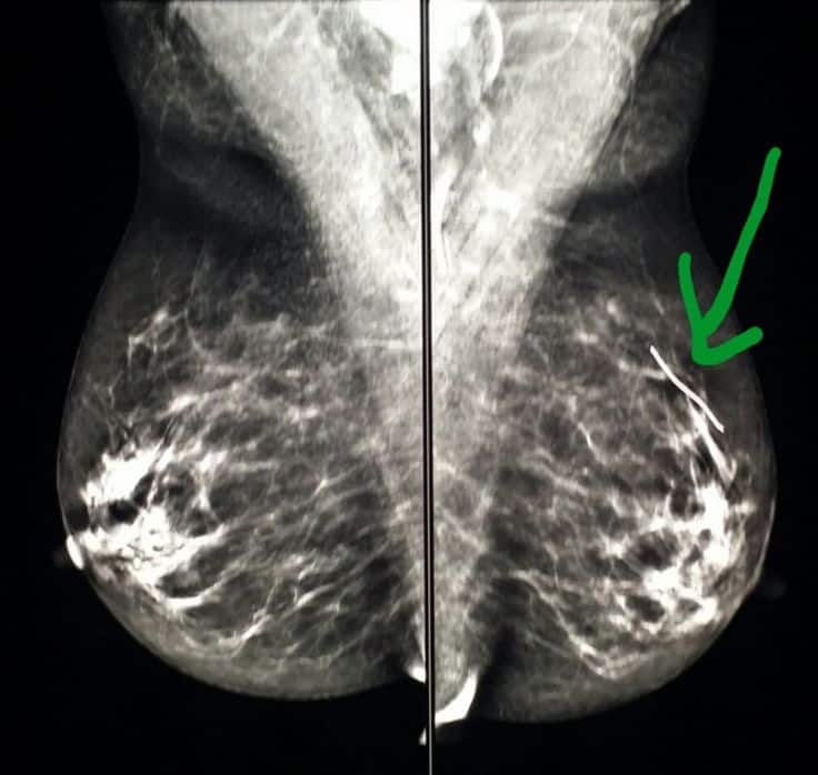
Breast Ultrasound Vs Mammogram: Image Quality
The quality of the images from a breast ultrasound and a mammogram are also different.
- Mammograms: If a screening mammogram identifies a suspicious area in the breast, you will likely need a diagnostic mammogram. A diagnostic mammogram takes more pictures than a routine screening mammogram and focuses on the affected area.
- Ultrasounds: A breast ultrasound cannot spotmicrocalcifications in the breast. Although calcifications are not always a sure sign of breast cancer, many early breast cancers are suspected because the calcifications are seen.
Recent studies suggest that people who have dense breasts could benefit from a mammogram plus fast breast magnetic resonance imaging . The combination of tests may produce fewer false positives than mammography and ultrasound alone.
In addition, fast breast MRI seems to be equal to conventional MRIs, which are the best test for finding breast cancer. Though they are good, they expensive and usually only offered to high-risk patients. Since fast breast MRI testing is relatively new, it is not available at every facility that offers breast cancer screenings.
How Accurate Is Ultrasound For Breast Cancer
The sensitivity and specificity of ultrasound for detecting breast carcinoma was 57.1% and 62.8% respectively with a positive predictive value of 68.1%, a negative predictive value of 99.5%, a positive likelihood ratio of 39 and a negative likelihood ratio of 0.07.
Can breast cancer be missed on mammogram and ultrasound?
About 20% to 30% of women with breast cancer have tumors that are missed by mammogram screening. And these interval breast cancers discovered between routine mammograms seem to be more lethal than those detected by screening.
What color is breast cancer on an ultrasound?
The images from a breast ultrasound are in black and white. Cysts, tumors, and growths will appear dark on the scan. However, just because there is a dark spot on your ultrasound, it does NOT mean that you have breast cancer.
Can you see cancer on an ultrasound?
Ultrasound cannot tell whether a tumor is cancer. Its use is also limited in some parts of the body because the sound waves cant go through air or through bone.
Determining The Nature Of A Breast Abnormality
The primary use of breast ultrasound is to help diagnose breast abnormalities, such as a lump, and to characterize potential abnormalities were seen in a mammogram or breast MRI. Ultrasound imaging can help to determine if an abnormality is:
- solid, which may be a non-cancerous lump of tissue or a cancerous tumor,
- fluid-filled, such as a benign cyst,
- or both cystic and solid.
In addition, Doppler ultrasound is used to assess the blood supply in breast lesions which can help determine the cause of the mass.
Recommended Reading: Do Breast Cancer Tumors Hurt
What Is An Ultrasound
Ultrasound screening is commonly used to study a developing fetus, heart and blood vessels, muscles, and tendons, and abdominal and pelvic organs. Beyond that, your radiologist would recommend an ultrasound scan when you get an abnormal mammogram.
The ultrasound uses high-frequency sound waves to look for tumors in the breast when they dont show up on X-rays .
In a breast ultrasound scan, your healthcare provider moves a wand-like device called a transducer over your breasts to get their images.
The transducer sends high-frequency sound waves that bounce off your breast tissue. It then picks up the bounced sound waves to develop pictures of the inside of your breasts.
What If Screening Uncovers A Suspicious Finding

Whether your screening includes mammography, ultrasound or MRIor some combination of all threefalse positive findings can occur. A false positive screening exam happens when the patient is called back for further workup of a finding that ultimately does not turn out to be cancer.
“The chances of having a false positive result is greater with a baseline mammogram. Studies show that women who have prior mammograms for comparison have fewer false positives,” Dr. Litwer says. These false findings are also more common among patients who:
- Are between the ages of 40 and 49
- Have dense breasts
You May Like: Can You Cure Breast Cancer Naturally
Supplemental Breast Cancer Screening
Mammography is the only screening tool for breast cancer that is confirmed to reduce deaths caused by breast cancer through early detection. However, mammograms do not detect all breast cancers. Some breast lesions and abnormalities are not visible or are difficult to interpret on mammograms. Breasts that are considered dense can make cancer harder to detect through typical breast exams. Many studies have shown that ultrasound and MRI can help supplement mammography by detecting breast cancers that may not be visible with mammography. Ultrasound can be offered as a screening tool for women who:
- Are at high risk for breast cancer and unable to undergo an MRI examination,
- are pregnant or should not be exposed to x-rays, which are necessary for a mammogram,
- have increased breast density when the breasts have a lot of glandular and connective tissue and not much fatty tissue.
What Are Dense Breasts
The breasts are made up of fatty, glandular, and connective tissues. Breast density is a measure of the amount of dense tissue compared to less dense tissue . Breasts that contain a higher proportion of glandular and connective tissues relative to fatty tissue are said to be dense.
Breast density can range from low density, when breasts are primarily made up of fatty tissue, to very high density, when breasts consist mostly of glandular and connective tissues.
The only way to determine a womans breast density is with a mammogram. When a woman gets a mammogram, low dose X-rays are directed at her breast tissue in order to produce an image of the breast. When doctors look at these images, they can distinguish between different tissues based on their appearance in the image.
Dense tissue, for instance, looks white in the image, while fatty tissue looks black. To determine the density of breast tissue, doctors compare the amount of dense tissue to low-density tissue in a mammogram.
You May Like: Is Breast Cancer In The Lymph Nodes Curable
Other Breast Cancer Imaging Options
Neither mammograms nor breast ultrasounds will find all breast cancers. There are options for people who are at a high risk for breast cancer.
- Breast magnetic resonance imaging uses a powerful magnetic and radio waves to generate highly detailed images, especially of the soft tissue. Breast MRI might be best for young people with dense breasts who have significant risk factors for breast cancer.
- Elastographymeasures the stiffness of breast tissue.
- Digital mammography uses less radiation than conventional mammograms.
- Optical mammography without compression uses infrared light instead of X-ray.
- Breast thermography can spot temperature variations suggestive of cancer but a 2016 study concluded that “thermography cannot substitute for mammography for the early diagnosis of breast cancer.”
These techniques continue to evolve as researchers look for better ways to find breast cancer in the earliest stages of the disease.
Breast Ultrasound Vs Diagnostic Mammogram
Is a breast ultrasound better than a mammogram? Though a doctor might order both as a follow-up after an abnormal screening mammogram, there are several notable differences in the procedures. Some distinctions between the two are as follows.
- The imaging modality: Modality is the type of imaging used during a diagnostic test. Ultrasound uses sound waves to create images, while x-rays use radiation from electromagnetic waves to capture an image.
- The quality of images produced: Ultrasound and x-rays produce different types of pictures. Generally speaking, ultrasound images cant capture microcalcifications. Those tiny calcium deposits can often be some of the earliest signs of breast cancer. However, a mammogram can detect them.
- The reasons for the imaging: A physician might order either an ultrasound or a diagnostic mammogram to follow up on an abnormal screening mammogram. A breast ultrasound has uses beyond being a follow-up tool. In some cases, a doctor will perform a breast biopsy, collecting a sample of tissue from the breast and testing it for cancer. They might use ultrasound imaging to guide the needle to the correct area of the breast.
Don’t Miss: How Would I Know If I Have Breast Cancer
Though Mammograms Can Miss Tumors They Provide More Information
A breast ultrasound or sonogram is not the same as a mammogram. Both are imaging tests that can be used to look for breast cancer. A mammogram is the gold standard for breast cancer detection, but a breast ultrasound can also identify specific changes in the breast.
Since each test complements the other, healthcare providers may use both breast ultrasounds and mammograms for breast cancer screening and diagnosis.
This article reviews the differences between breast ultrasounds and mammograms. You will learn about the benefits, limitations, and risks of each type of imaging test as well as other options for diagnosing breast cancer.
Additional Information For Webmds Visitors From The European Economic Area
When you use the Services, we collect, store, use and otherwise process your personal information as described in this Privacy Policy. We rely on a number of legal bases to process your information, including where: necessary for our legitimate interests in providing and improving the Services including offering you content and advertising that may be of interest to you necessary for our legitimate interest in keeping the Services, Sites and Apps safe and secure necessary for the legitimate interests of our service providers and partners necessary to perform our contractual obligations in the WebMD Terms of Use you have consented to the processing, which you can revoke at any time you have expressly made the information public, e.g., in public forums necessary to comply with a legal obligation such as a law, regulation, search warrant, subpoena or court order or to exercise or defend legal claims and necessary to protect your vital interests, or those of others.
Where we process your personal information for direct marketing purposes, you can opt-out through the unsubscribe link in the email communications we send to you, by changing your subscription preferences in your account settings or as otherwise specified in this Privacy Policy.
Also Check: How Do I Get Checked For Breast Cancer
How Is The Procedure Performed
You will lie on your back or on your side on the exam table. The sonographer may ask you to raise your arm above your head.
The radiologist or sonographer will position you on the exam table. They will apply a water-based gel to the area of the body under examination. The gel will help the transducer make secure contact with the body. It also eliminates air pockets between the transducer and the skin that can block the sound waves from passing into your body. The sonographer places the transducer on the body and moves it back and forth over the area of interest until it captures the desired images.
There is usually no discomfort from pressure as they press the transducer against the area being examined. However, if the area is tender, you may feel pressure or minor pain from the transducer.
Doctors perform Doppler sonography with the same transducer.
Once the imaging is complete, the technologist will wipe off the clear ultrasound gel from your skin. Any portions that remain will dry quickly. The ultrasound gel does not usually stain or discolor clothing.
When Is A Breast Ultrasound Needed

Typically, healthcare providers dont use breast ultrasound on its own to screen for breast cancer. More often, they recommend an ultrasound to follow up on suspicious areas seen on a mammogram. Because hand-held ultrasound uses a small probe to check the tissue, it is most useful when there is a specific targeted area of interest within the breast to examine. Mammography is still the best tool for screening the entire breast, even indense breasts.
A healthcare provider may recommend a breast ultrasound for many different reasons. Some of the most common are:
- Checking if a breast lump is a fluid-filled breast cyst or a solid mass .
- Investigating a focal area in the breast that appeared abnormal on a mammogram.
- Examining a pregnant womans breasts in conjunction with physical exam. Occasionally, a mammogram is also used in pregnant women because radiation doses are very low and the abdomen can be shielded if concern for breast cancer detection is high.
- Guiding a needle into a mass to sample tissue for a biopsy. Pathologists can then evaluate the tissue under a microscope to determine if the mass is breast cancer.
Recommended Reading: Do Larger Breasts Increase Cancer Risk
Why Might I Need A Breast Ultrasound
A breast ultrasound is most often done to find out if a problem found by a mammogram or physical exam of the breast may be a cyst filled with fluid or a solid tumor.
Breast ultrasound is not usually done to screen for breast cancer. This is because it may miss some early signs of cancer. An example of early signs that may not show up on ultrasound are tiny calcium deposits called microcalcifications.
Ultrasound may be used if you:
-
Have particularly dense breast tissue. A mammogram may not be able to see through the tissue.
-
Are pregnant. Mammography uses radiation, but ultrasound does not. This makes it safer for the fetus.
-
Are younger than age 25
Your healthcare provider may also use ultrasound to look at nearby lymph nodes, help guide a needle during a biopsy, or to remove fluid from a cyst.
Your healthcare provider may have other reasons to recommend a breast ultrasound.
What Is A Mammogram
Mammography is a breast imaging procedure that gives an X-ray image of your breasts. Regular mammograms are the best way for your oncologist to find breast cancer at an early stage.
Sometimes, a mammogram can help detect breast cancer up to three years before you feel a cyst, lesion, or any abnormal change in your breast tissue.
During a 2D mammogram, youll stand in front of a special X-ray machine. Your radiologist will place your breast on a plastic plate while another plate will firmly press your breast from above.
These plates flatten the breast and keep it still while taking the X-ray. After getting the top and bottom X-rays of your breast, your radiologist will take the side view as well. Its common to feel some pressure during a mammogram.
A 3D mammogram follows this procedureexcept that instead of creating only four images from two angles, 3D mammography produces several images from all possible angles to create a three-dimensional model of your breast.
Again, the mammography procedure occurs in two phasesa screening mammography and a diagnostic mammography.
In the former, your oncologist tries to detect breast changes in women without symptoms or new breast abnormalities. And with a diagnostic mammogram, your oncologist investigates the suspicious breast changes.
You May Like: Can You Live With Stage 4 Breast Cancer
How Fast Do Breast Tumors Grow
Studies show that even though breast cancer happens more often now than it did in the past, it doesnt grow any faster than it did decades ago. On average, breast cancers double in size every 180 days, or about every 6 months. Still, the rate of growth for any specific cancer will depend on many factors.
When Is Breast Ultrasound Used
Ultrasound is not typically used as a routine screening test for breast cancer. But it can be useful for looking at some breast changes, such as lumps . Ultrasound can be especially helpful in women with dense breast tissue, which can make it hard to see abnormal areas on mammograms. It also can be used to get a better look at a suspicious area that was seen on a mammogram.
Ultrasound is useful because it can often tell the difference between fluid-filled masses like cysts and solid masses .
Ultrasound can also be used to help guide a biopsy needle into an area of the breast so that cells can be taken out and tested for cancer. This can also be done in swollen lymph nodes under the arm.
Ultrasound is widely available and is fairly easy to have done, and it does not expose a person to radiation. It also tends to cost less than other testing options.
Also Check: 8 Cycles Of Chemotherapy For Breast Cancer
Why Is It Important For Women To Know If They Have Dense Breasts
There are two reasons why this information is important.
- Dense breast tissue can make it harder for doctors to detect small tumors in mammograms. Like dense breast tissue, tumors look white in mammogram images. This makes it more difficult for doctors to distinguish tumors from dense breast tissue. Because of this, for women who have dense breast tissue, mammograms may overlook tumors.
- Women who have dense breasts have a higher-than-average risk of developing breast cancer. This risk rises with increasing breast density.
Its important to note that if women with dense breasts are diagnosed with breast cancer, they are not at higher risk of dying from breast cancer, compared to women without dense breasts.
Purpose Of Breast Ultrasounds Vs Mammograms
A mammogram is an X-ray of the breasts. Mammograms are the most effective breast cancer screening test. They can take multiple pictures of the breast and identify calcifications . In addition, mammograms are important for diagnosing and following up after breast cancer.
A breast ultrasound or sonogram is generally used for diagnostic reasons. For example, an ultrasound is most helpful when evaluating dense breasts or a suspicious lump found on a mammogram.
A breast ultrasound is good at distinguishing a benign fluid-filled cyst from a solid mass. An ultrasound of the breast can help define a mass found by touch, even if it does not appear on a mammogram.
You May Like: What Percentage Survive Breast Cancer