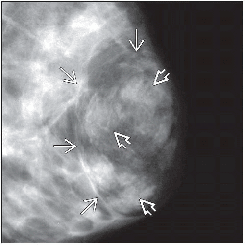
Other Potential Signs Of Breast Cancer Include:
- Lump in the breast or in the underarm
- A spontaneous or bloody discharge from the nipple
- New retraction or indentation of the nipple
- A change in the size or contour of the breast
- Any flattening or indentation of the skin over the breast
- Redness or pitting of the skin over the breast, like the skin of an orange
- Crustiness, ulceration or rash of the nipple or areola
A number of conditions other than breast cancer can cause breasts to change in size or feel. Breast tissue changes naturally during pregnancy and a womans menstrual cycle. Other possible causes of non-cancerous breast changes include fibrocystic changes, cysts, fibroadenomas, infection or injury.
If you find a lump or other change in your breast even if a recent mammogram was normal you should call us immediately. If you havent yet gone through menopause, you may want to wait through one menstrual cycle before seeing your doctor. If the change hasnt gone away after a month, have it evaluated.
Checking The Lymph Nodes
The usual treatment is surgery to remove the cancer. Before your surgery you have an ultrasound scan to check the lymph nodes in the armpit close to the breast. This is to see if they contain cancer cells. If breast cancer spreads, it usually first spreads to the lymph nodes close to the breast.
Depending on the results of your scan you might have:
- a sentinel lymph node biopsy during your breast cancer operation
- surgery to remove your lymph nodes
You may have other treatments after surgery.
How Is The Stage Determined
The staging system most often used for breast cancer is the American Joint Committee on Cancer TNM system. The most recent AJCC system, effective January 2018, has both clinical and pathologic staging systems for breast cancer:
- The pathologic stage is determined by examining tissue removed during an operation.
- Sometimes, if surgery is not possible right away or at all, the cancer will be given a clinical stage instead. This is based on the results of a physical exam, biopsy, and imaging tests. The clinical stage is used to help plan treatment. Sometimes, though, the cancer has spread further than the clinical stage estimates, and may not predict the patients outlook as accurately as a pathologic stage.
In both staging systems, 7 key pieces of information are used:
- The extent of the tumor : How large is the cancer? Has it grown into nearby areas?
- The spread to nearby lymph nodes : Has the cancer spread to nearby lymph nodes? If so, how many?
- The spread to distant sites : Has the cancer spread to distant organs such as the lungs or liver?
- Estrogen Receptor status: Does the cancer have the protein called an estrogen receptor?
- Progesterone Receptor status: Does the cancer have the protein called a progesterone receptor?
- HER2 status: Does the cancer make too much of a protein called HER2?
- Grade of the cancer : How much do the cancer cells look like normal cells?
In addition, Oncotype Dx® Recurrence Score results may also be considered in the stage in certain situations.
Don’t Miss: Baking Soda For Breast Cancer
Understanding Tumor Size Measurement
Tumor size is typically measured using millimeters or centimeters. For example, a tumor could be 2 centimeters or 20 millimeters. Common everyday foods may help with understanding measurements:
- 1 cm is about the width of a pea
- 2 cm is about the size of a peanut
- 3 cm is about the size of a grape
- 4 cm is about the size of a walnut
- 5 cm is about the size of a lime
- 6 cm is about the size of an egg
- 7 cm is about the size of a peach
- 10 cm is about the size of a grapefruit
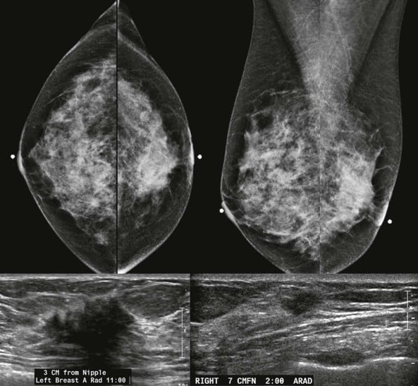
Tumor size is measured based on an imaging scan or surgical removal of the tumor. Tests such as mammograms or ultrasounds may be used to take images and measure the tumor, and often may be instrumental in first detecting the tumor. If the tumor is operable, meaning it may be safely removed during surgery, it will also be measured after removal. Tumors are measured at their widest point.
Influence Of Tumour Stage At Breast Cancer Detection On Survival In Modern Times: Population Based Study In 173 797 Patients

- Accepted 4 September 2015
Also Check: How To Treat Stage 0 Breast Cancer
What Is Stage Iv Breast Cancer
Stage IV is the most advanced stage of breast cancer. It has spread to nearby lymph nodes and to distant parts of the body beyond the breast. This means it possibly involves your organs such as the lungs, liver, or brain or your bones.
Breast cancer may be stage IV when it is first diagnosed, or it can be a recurrence of a previous breast cancer that has spread.
Starting With Neoadjuvant Therapy
Most often, these cancers are treated with neoadjuvant chemotherapy. For HER2-positive tumors, the targeted drug trastuzumab is given as well, often along with pertuzumab . This may shrink the tumor enough for a woman to have breast-conserving surgery . If the tumor doesnt shrink enough, a mastectomy is done. Nearby lymph nodes will also need to be checked. A sentinel lymph node biopsy is often not an option for stage III cancers, so an axillary lymph node dissection is usually done.
Often, radiation therapy is needed after surgery. If breast reconstruction is planned, it is usually delayed until after radiation therapy is done. For some, additional chemo is given after surgery as well.
After surgery, some women with HER2-positive cancers will be treated with trastuzumab for up to a year. Many women with HER2-positive cancers will be treated first with trastuzumab followed by surgery and then more trastuzumab for up to a year. If after neoadjuvant therapy, any residual cancer is found at the time of surgery, ado-trastuzumab emtansine may be used instead of trastuzumab. It is given every 3 weeks for 14 doses. For women with hormone receptor-positive cancer that is in the lymph nodes, who have completed a year of trastuzumab, the doctor might also recommend additional treatment with an oral targeted drug called neratinib for a year.
Recommended Reading: What Does Stage 1 Breast Cancer Mean
Putting It All Together
All of the TNM information will be combined twice, once by the surgeon and again by the pathologist . Each expert will give an opinion about your case in terms of its TNM stage. To officially determine the breast cancer stage, your team may need to know more about:
- Cancer cells have not spread out of the breast into the lymph nodes.
Stage 1B:
- A small group of cancer cells measuring between 0.2 millimeters and 2 mm is found in the lymph nodes.
- A stage 1A tumor may or may not exist.
What Is A Histological Work
Determining your type of breast cancer begins with a histological workup, a summary prepared by the pathologist after you undergo a biopsy. Essentially, the histological evaluation is the microscopic analysis of the chemical and cellular properties associated with a suspicious breast tumor. The pathologists here at Providence Saint Johns will also confirm the size of the breast tumor where necessary for breast cancer staging purposes. The histological evaluation is essential to determine the most effective treatment recommendations following surgery.
Read Also: Why Would You Do Chemo Before Surgery For Breast Cancer
How Is Breast Cancer Treated
There are several breast cancer treatment options, including surgery, chemotherapy, radiation therapy, hormone therapy, immunotherapy and targeted drug therapy. Whats right for you depends on many factors, including the location and size of the tumor, the results of your lab tests and whether the cancer has spread to other parts of your body. Your healthcare provider will tailor your treatment plan according to your unique needs. Its not uncommon to receive a combination of different treatments, too.
Breast cancer surgery
Breast cancer surgery involves removing the cancerous portion of your breast and an area of normal tissue surrounding the tumor. There are different types of surgery depending on your situation, including:
Chemotherapy for breast cancer
Your healthcare provider may recommend chemotherapy for breast cancer before a lumpectomy in an effort to shrink the tumor. Sometimes, its given after surgery to kill any remaining cancer cells and reduce the risk of recurrence . If the cancer has spread beyond your breast to other parts of your body, then your healthcare provider may recommend chemotherapy as a primary treatment.
Radiation therapy for breast cancer
Radiation therapy for breast cancer is typically given after a lumpectomy or mastectomy to kill remaining cancer cells. It can also be used to treat individual metastatic tumors that are causing pain or other problems.
Hormone therapy for breast cancer
Immunotherapy for breast cancer
Rfs And Efs In Women With Er
When the data from the groups of women in B-06, B-13, and B-19 who received no systemic therapy were combined, the 8-year RFS was 81%. When the data from women who received chemotherapy in the three studies were combined, the 8-year RFS was 90% . The cumulative probabilities of events comprising EFS were computed for each treatment group . In women treated with surgery alone, the cumulative incidence of all events was 30%. There was an 18% probability of recurrence at local-regional or distant sites through 8 years 10% of these events were distant recurrences, 3% were IBTR, and 5% were recurrences at other local-regional sites. The cumulative incidence of second cancer in the contralateral breast was 5% the cumulative incidence of other second primary cancers was 7%. There were no deaths before recurrence or second cancer as a first event . In patients who received postoperative chemotherapy, the cumulative incidence of all events was 18% through 8 years 9% of the total cumulative incidence was from local-regional recurrences or distant recurrences , 4% was from cancers in the contralateral breast, 3% was from second cancers other than breast cancer, and 2% was from deaths with no evidence of disease . The frequency of IBTR after lumpectomy and breast irradiation decreased from 8% to 3% as a result of chemotherapy .
Recommended Reading: How Is Breast Cancer Caused
Er And Progesterone Receptor Determinations
The U.S. Food and Drug Administration approved the use of the enzyme immunoassay kit from Abbott Laboratories for analysis of ER and PgR in October 1988 and September 1990, respectively. The NSABP then allowed these methods to be used in their trials for determining receptor status in patients who had 24 mm3 of biopsy tissue that weighed 100200 mg. Later, it was reported that the enzyme immunoassay procedures were highly reproducible in breast carcinoma biopsies of less than 100 mg of tissue taken from patients during the late 1980s and early 1990s.
Invasive Ductal Carcinoma Stages

Invasive ductal carcinoma stages provide physicians with a uniform way to describe how far a patients cancer may have spread beyond its original location in a milk duct. This information can be helpful when evaluating treatment options, but it is not a prognostic indicator in and of itself. Many factors can influence a patients outcome, so the best source of information for understanding a breast cancer prognosis is always a physician who is familiar with the patients case.
Don’t Miss: Does Breast Pain Indicate Cancer
Stage Groups For Breast Cancer
Doctors assign the stage of the cancer by combining the T, N, and M classifications , the tumor grade, and the results of ER/PR and HER2 testing. This information is used to help determine your prognosis . The simpler approach to explaining the stage of breast cancer is to use the T, N, and M classifications alone. This is the approach used below to describe the different stages.
Most patients are anxious to learn the exact stage of the cancer. If you have surgery as the first treatment for your cancer, your doctor will generally confirm the stage of the cancer when the testing after surgery is finalized, usually about 5 to 7 days after surgery. When systemic treatment is given before surgery, which is typically with medications and is called neoadjuvant therapy, the stage of the cancer is primarily determined clinically. Doctors may refer to stage I to stage IIA cancer as “early stage” and stage IIB to stage III as “locally advanced.” Stage 0: Stage zero describes disease that is only in the ducts of the breast tissue and has not spread to the surrounding tissue of the breast. It is also called non-invasive or in situ cancer . Stage IA: The tumor is small, invasive, and has not spread to the lymph nodes . Stage IB: Cancer has spread to the lymph nodes and the cancer in the lymph node is larger than 0.2 mm but less than 2 mm in size. There is either no evidence of a tumor in the breast or the tumor in the breast is 20 mm or smaller .
Stage IIA: Any 1 of these conditions:
N Categories For Breast Cancer
N followed by a number from 0 to 3 indicates whether the cancer has spread to lymph nodes near the breast and, if so, how many lymph nodes are involved.
Lymph node staging for breast cancer is based on how the nodes look under the microscope, and has changed as technology has gotten better. Newer methods have made it possible to find smaller and smaller groups of cancer cells, but experts haven’t been sure how much these tiny deposits of cancer cells influence outlook.
Its not yet clear how much cancer in the lymph node is needed to see a change in outlook or treatment. This is still being studied, but for now, a deposit of cancer cells must contain at least 200 cells or be at least 0.2 mm across for it to change the N stage. An area of cancer spread that is smaller than 0.2 mm doesn’t change the stage, but is recorded with abbreviations that indicate the type of special test used to find the spread.
If the area of cancer spread is at least 0.2 mm , but still not larger than 2 mm, it is called a micrometastasis . Micrometastases are counted only if there aren’t any larger areas of cancer spread. Areas of cancer spread larger than 2 mm are known to influence outlook and do change the N stage. These larger areas are sometimes called macrometastases, but are more often just called metastases.
NX: Nearby lymph nodes cannot be assessed .
N0: Cancer has not spread to nearby lymph nodes.
N1c: Both N1a and N1b apply.
N3: Any of the following:
N3a: either:
N3b: either:
Don’t Miss: How To Lose Weight After Breast Cancer
What Is The Survival Rate For Invasive Ductal Carcinoma
The survival rate for this malignancy varies depending on the stage the patient is at. For example:
- If invasive ductal carcinoma has not spread beyond the breast, the five-year survival rate is approximately 99%.
- If the cancer has spread to nearby structures or lymph nodes, the five-year survival rate is approximately 86%.
- If the malignancy has spread to a distant area of the body, the five-year survival rate is approximately 27%.
When considering these numbers, its important to remember that they are just general benchmarks and should not be used to predict a specific persons chances of survival. There are a number of factors that can influence a patients prognosis, such as:
- Whether the cancer is new or recurring
- How far the cancer had progressed by the time it was diagnosed
- The cancers hormone-receptor status and HER2 status
- How quickly the cancer cells are growing
- How the cancer is responding to treatment
- The patients age, menopausal status and overall health
Plus, reported survival rates will of course be dated, and as newer treatment methods are developed, these rates will likely improve.
T Categories For Breast Cancer
T followed by a number from 0 to 4 describes the main tumor’s size and if it has spread to the skin or to the chest wall under the breast. Higher T numbers mean a larger tumor and/or wider spread to tissues near the breast.
TX: Primary tumor cannot be assessed.
T0: No evidence of primary tumor.
Tis: Carcinoma in situ
T1 : Tumor is 2 cm or less across.
T2: Tumor is more than 2 cm but not more than 5 cm across.
T3: Tumor is more than 5 cm across.
T4 : Tumor of any size growing into the chest wall or skin. This includes inflammatory breast cancer.
Don’t Miss: Signs And Symptoms Of Metastatic Breast Cancer
Stages Of Breast Cancer
Your breast cancer stage indicates the severity of the disease upon diagnosis. Your breast cancer stage indicates the severity of the disease upon diagnosis. Your cancer stage will always stay the same, even if the cancer shrinks or spreads during or after treatment. For instance, if youre diagnosed with stage 1 breast cancer, but the tumor later grows and spreads, its not considered stage 3 or 4 breast cancer. To determine whether the cancer has responded to treatment, a new stage may later be assigned an r in front of it to show that its different from the original stage.
Breast cancer staging is classified by:
- The size and location of the tumor
- Whether the cancer has spread to nearby lymph nodes or other parts of the body
- The grade of the tumoror how likely it is to grow and spread
- Whether certain biomarkershormone receptors or other proteinshave been found
All these attributes help your care team determine how to treat your cancer.
To assess the location, size and spread of cancer, your care team will use the TNM Staging System, developed and updated for breast cancer by the American Joint Committee on Cancer .
- TNM stands for Tumor-Node-Metastasis, which are important factors in determining the severity of your cancer.
- All cancers may be evaluated by TNM markers, but breast cancer staging also uses a few extra criteria for a more detailed description.
- Ultimately, your specific combination of TNM and these other markers will determine your cancers stage.